Key Takeaways
- Clinical Bottom Line
- Why OTSC deserves a fresh look
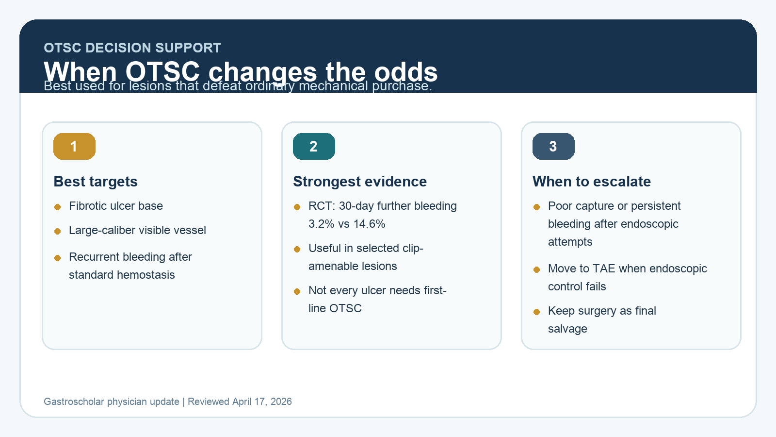
- The lesions where OTSC makes the most sense
- The current evidence is stronger than it was even 3 years ago
Clinical Bottom Line
| Clinical question | 2026 answer |
|---|---|
| When should OTSC enter the conversation? | When standard clips or thermal therapy are likely to fail because the lesion is fibrotic, large-vessel, awkward, recurrent, or technically unforgiving. |
| Is OTSC only rescue therapy? | No. It remains a rescue option, but randomized data now support first-line use in selected clip-amenable nonvariceal upper GI bleeding lesions. |
| What is the best-quality trial signal? | In a 190-patient randomized trial, 30-day further bleeding was 3.2% with OTSC versus 14.6% with standard endoscopic therapy. |
| Does OTSC replace core ulcer-bleeding care? | No. Resuscitation, lesion classification, post-hemostasis PPI therapy, H. pylori management, and a plan for rebleeding still determine outcomes. |
| When do you move beyond OTSC? | If the lesion cannot be meaningfully captured or bleeding persists despite endoscopic options, move to transcatheter arterial embolization and keep surgery in reserve. |
Why OTSC deserves a fresh look
OTSC is powerful, but the real clinical question is where it improves the probability of durable hemostasis enough to justify the cap exchange, deployment planning, and cost. The strongest use cases are lesions where tissue capture and vessel compression are likely to outperform standard clips or contact thermal therapy.
The lesions where OTSC makes the most sense
- Bleeding peptic ulcers with a fibrotic or indurated base where through-the-scope clips will not grab enough tissue.
- Large-caliber nonbleeding visible vessels or active spurting lesions where definitive compression matters more than temporary blanching.
- Dieulafoy lesions and selected post-intervention defects where deep tissue capture is achievable.
- Recurrent bleeding after previously successful standard hemostasis.
What OTSC does well is not just “clip harder.” It captures more tissue, generates a more durable mechanical seal, and can stabilize lesions that repeatedly defeat standard clips or thermal therapy.
The current evidence is stronger than it was even 3 years ago
The most useful randomized signal comes from the 2023 multicenter Annals trial comparing OTSC with standard endoscopic treatment as initial therapy in nonvariceal upper GI bleeding. Among 190 adults with active bleeding or a nonbleeding visible vessel, 30-day further bleeding was 3.2% in the OTSC group versus 14.6% with standard therapy. That result is hard to ignore.
Systematic reviews and meta-analyses of randomized and comparative studies have largely pointed in the same direction: lower rebleeding and less need for rescue intervention in appropriately selected lesions. The key phrase is appropriately selected. These data do not mean every peptic ulcer should get an OTSC first.
What still makes standard therapy reasonable
For many lesions, standard therapy remains entirely appropriate.
- Small focal vessels in nonfibrotic tissue may be treated well with TTS clips or contact thermal therapy.
- Epinephrine remains useful to improve visualization, but not as stand-alone treatment.
- Topical hemostatic powder remains helpful when visualization is poor or the bleeding field is diffuse, especially as salvage or bridge therapy.
OTSC becomes more attractive as the lesion becomes less forgiving, not simply because the bleeding looks dramatic.
Practical deployment mistakes that still cause failure
| Failure mode | Why OTSC underperforms |
|---|---|
| Tangential approach | If the cap cannot face the lesion squarely, tissue capture becomes incomplete and vessel compression is unreliable. |
| Huge cavitary ulcer | The vessel may sit deep in a crater that is difficult to suction adequately into the cap. |
| Inadequate lesion preparation | If the field is not cleared and the target is not defined, the clip may capture the wrong tissue and create false reassurance. |
| No escalation plan | Time is lost if OTSC is treated as the final answer rather than one step in a structured refractory-bleeding pathway. |
Where OTSC fits in the current guideline ecosystem
ACG still positions over-the-scope clips mainly as a preferred option for recurrent ulcer bleeding after prior successful hemostasis. ESGE 2021 supports cap-mounted clips for persistent bleeding refractory to standard modalities. The 2023 randomized data move the field closer to selected first-line OTSC use, especially in lesions where the endoscopist already suspects standard therapy is mechanically underpowered.
Do not forget the rest of the bleeding pathway
- Resuscitate first.
- Perform endoscopy within 24 hours after stabilization.
- Give high-dose PPI therapy for 72 hours after successful hemostasis of a high-risk ulcer.
- Test and treat for H. pylori where relevant.
- If bleeding recurs, repeat endoscopy is usually the next step.
- If endoscopic control fails, move to transcatheter arterial embolization. Keep surgery as the final salvage pathway.
Selected references
- Barkun A, et al. OTSC versus standard endoscopic treatment as initial therapy in nonvariceal upper GI bleeding. Ann Intern Med. 2023.
- STING-2 randomized trial of OTSC versus standard treatment in high-risk acute nonvariceal upper GI bleeding.
- Systematic review and meta-analysis of OTSC versus standard first-line therapy in non-variceal upper GI bleeding. 2023.
- ACG Clinical Guideline: Upper Gastrointestinal and Ulcer Bleeding. 2021.
- ESGE Guideline Update: Nonvariceal Upper Gastrointestinal Hemorrhage. 2021.
Last reviewed April 17, 2026. This update is written for clinicians choosing between standard endoscopic hemostasis, OTSC, and escalation pathways in upper GI bleeding.