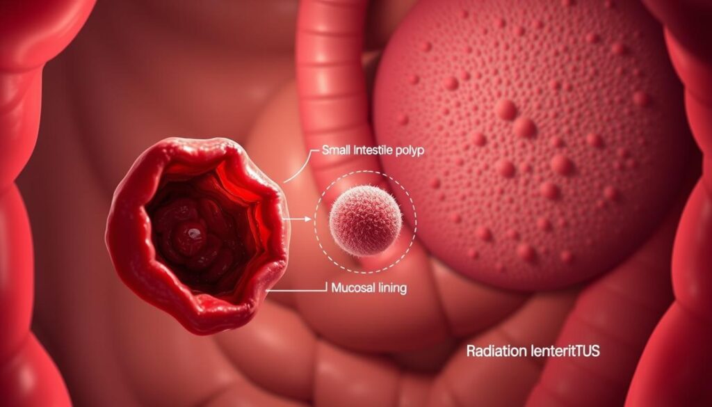
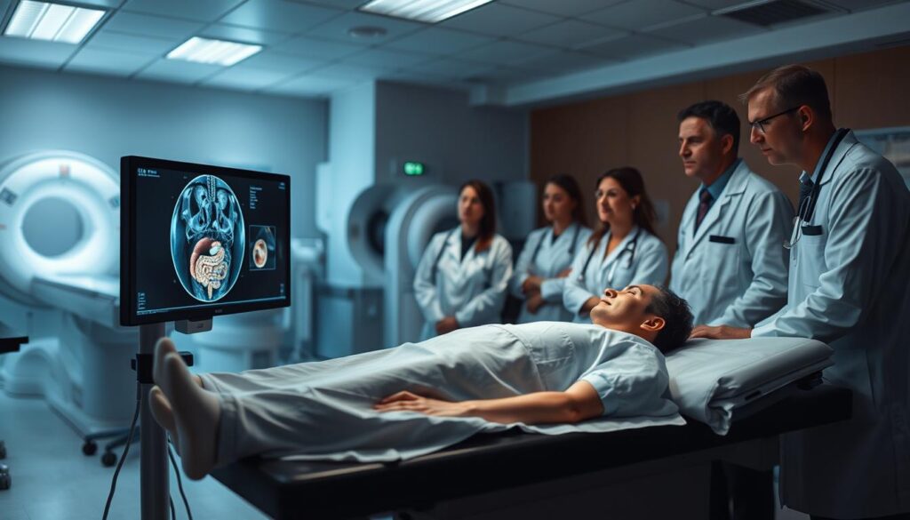
Video capsule endoscopy has become a cornerstone in diagnosing gastrointestinal bleeding, particularly in the small intestine. This non-invasive technique allows clinicians to visualize areas that are difficult to access with traditional methods. Studies show a diagnostic yield ranging from 43.5% to 90%, depending on the clinical presentation1.
FDA-approved systems like PillCam SB, Endocapsule, and MiroCam are widely used for this purpose. These devices have demonstrated a pooled detection rate of 59.4% across 227 studies, making them invaluable in clinical practice1. The completion rate for these procedures is notably high, with rates of 88.1% for overt cases and 91.5% for occult cases2.
Despite its effectiveness, the procedure carries a minimal risk of capsule retention, ranging from 0.7% to 1.5% in patients with obscure gastrointestinal bleeding2. Complementary use with CT enterography further enhances its diagnostic capabilities, particularly for detecting masses and other structural abnormalities.
Key Takeaways
- Video capsule endoscopy is a non-invasive method for diagnosing gastrointestinal bleeding.
- Diagnostic yield varies from 43.5% to 90%, depending on clinical factors1.
- FDA-approved systems include PillCam SB, Endocapsule, and MiroCam.
- Capsule retention risk is low, ranging from 0.7% to 1.5%2.
- CT enterography complements capsule endoscopy for mass detection.
Understanding Small Bowel Bleeding and Its Clinical Significance
Small bowel bleeding presents unique diagnostic challenges due to its complex anatomy. The small intestine, spanning 5-8 meters, has overlapping loops and mobile positioning, making it difficult to identify bleeding sources3. This complexity often delays accurate diagnosis and treatment.
Why Small Bowel Bleeding Challenges Diagnosis
The small intestine’s length and intricate structure hinder traditional diagnostic methods. Overlapping loops and mobility often obscure lesions, leading to missed findings. Studies show that 30% of iron deficiency anemia cases remain unexplained after initial evaluations3.
Common findings include angioectasias (50%), inflammation (26.8%), and tumors (8.8%)4. These lesions are often subtle and require advanced imaging techniques for detection.
Differences Between Overt and Occult Bleeding
Overt bleeding presents with visible signs like melena or hematochezia, while occult bleeding is associated with iron deficiency anemia. The diagnostic yield for overt cases is 66.6%, compared to 25.7% for occult cases3.
Proximal small bowel lesions are more common in patients presenting with melena. This distinction is crucial for guiding diagnostic strategies and improving outcomes.
| Type of Bleeding | Presentation | Diagnostic Yield |
|---|---|---|
| Overt | Melena/Hematochezia | 66.6% |
| Occult | Iron Deficiency Anemia | 25.7% |
Understanding these differences helps clinicians tailor diagnostic approaches and improve diagnostic yield. Accurate identification of bleeding sources is essential for effective treatment and patient outcomes.
Capsule Endoscopy Interpretation Guidelines for Small Bowel Bleeding
Interpreting findings from video-based diagnostic tools requires a systematic approach to ensure accuracy. Standardized reporting protocols, such as the SAVER framework, enhance consistency in identifying lesions. These protocols include SPICE, Saurin, and Lewis scores, which provide structured criteria for evaluation5.
Dual-frame review at 6-8 frames per second is recommended for the proximal small bowel. This technique improves lesion detection by capturing subtle abnormalities that might be missed at lower frame rates1. Second-reader verification is also advised for negative studies to minimize oversight.
A systematic review of the entire gastrointestinal tract, including stomach and colon segments, ensures comprehensive evaluation. Correlation with CT enterography (CTE) findings is essential for confirming masses and other structural abnormalities5.
“The integration of advanced imaging techniques with standardized protocols significantly improves diagnostic accuracy.”
Studies highlight the importance of dynamic contrast-enhanced CT in detecting active bleeding, which complements findings from video-based tools5. This dual approach enhances the overall diagnostic workflow.
| Review Technique | Purpose | Benefit |
|---|---|---|
| Dual-Frame Review | Proximal Small Bowel | Improved Lesion Detection |
| Second-Reader Verification | Negative Studies | Reduced Oversight |
| Systematic GI Tract Review | Comprehensive Evaluation | Enhanced Accuracy |
These guidelines ensure a methodical approach to interpreting findings, improving outcomes for patients with gastrointestinal bleeding. The combination of advanced imaging and structured protocols sets a new standard in diagnostic accuracy1.
Indications for Capsule Endoscopy in Suspected Small Bowel Bleeding
Identifying the source of gastrointestinal bleeding often requires advanced diagnostic tools. These tools are particularly useful when traditional methods, such as upper and lower endoscopic examinations, yield inconclusive results6. In such cases, advanced imaging techniques become essential for accurate diagnosis.
When to Choose Advanced Imaging Over Other Tests
Advanced diagnostic tools are preferred when initial endoscopic studies fail to identify the bleeding source. For example, they show a diagnostic yield of 72.5% compared to 48.7% for push enteroscopy in randomized controlled trials6. This makes them a first-line option after negative bidirectional endoscopy.
Additionally, these tools outperform angiography, with a yield of 53.3% versus 20%4. Their non-invasive nature and ability to visualize hard-to-reach areas further enhance their clinical utility.
Patient Groups with Higher Diagnostic Yield
Certain patient groups benefit more from advanced diagnostic tools. Hospitalized individuals and those on anticoagulation therapy often show higher diagnostic yield4. Early deployment within three days of hospitalization can also reduce the duration of hospital stays.
Patients with connective tissue diseases are another high-yield group, with an odds ratio of 2.24 for positive findings4. These insights help clinicians tailor diagnostic strategies for better outcomes.
How Capsule Endoscopy Works: A Step-by-Step Overview
The process of video capsule endoscopy involves a detailed and systematic approach to capturing images of the gastrointestinal tract. Patients are required to fast for 12 hours before ingestion to ensure optimal imaging conditions7. This preparation minimizes obstructions and enhances the clarity of the captured images.
Once ingested, the capsule passively moves through the digestive system, capturing high-quality images at an adaptive frame rate8. Dual-camera systems are used to ensure comprehensive coverage, capturing 2-6 frames per second depending on the capsule’s movement9. This technology provides natural tissue colors and balanced illumination for accurate diagnosis.
The procedure typically takes 8 to 10 hours, with a small bowel transit time ranging from 1.5 to 5 hours7. During this period, the capsule transmits images to an external receiver worn by the patient. Real-time viewer verification ensures the capsule has passed the pyloric region, confirming proper placement8.
After the procedure, the collected data is reviewed using proprietary software such as QuickView and TOP 100. These tools enable rapid screening and efficient video compilation, reducing review time by up to 40%9. The software also includes an enhanced GI map and atlas for better visualization and comparison of pathologies.
| Step | Details |
|---|---|
| Preparation | 12-hour fasting period |
| Ingestion | Capsule equipped with dual cameras |
| Transit | Passive movement through the digestive system |
| Imaging | Adaptive frame rate, 2-6 frames/sec |
| Review | Proprietary software for efficient analysis |
Data encryption and HIPAA-compliant storage ensure patient privacy and security throughout the process. This step-by-step approach makes small-bowel capsule endoscopy a reliable and efficient diagnostic tool for gastrointestinal conditions.
Preparing Patients for Capsule Endoscopy
Proper preparation is essential for ensuring the success of diagnostic procedures. This involves dietary adjustments, medication management, and assessing potential risks. These steps optimize the accuracy and safety of the process.
Dietary and Medication Adjustments
Patients are advised to follow a 24-hour clear liquid diet before the procedure. This ensures optimal imaging conditions by minimizing obstructions. Additionally, nonsteroidal anti-inflammatory drugs (NSAIDs) should be discontinued seven days prior to reduce the risk of mucosal irritation10.
For enhanced bowel preparation, a compound polyethylene glycol electrolyte solution is administered at specific intervals. This protocol improves visualization and diagnostic yield10.
Assessing Risk of Retention
The risk of retention varies based on clinical factors. High-risk groups include patients with Crohn’s disease, prior radiation, or surgical history. CT enterography is recommended for stricture detection in these cases11.
Patency capsules are used to evaluate gastrointestinal tract patency before the procedure. This reduces retention rates in high-risk patients, ensuring safer outcomes4.
“Rigorous preparation protocols significantly enhance diagnostic accuracy and patient safety.”
By addressing dietary, medication, and risk factors, clinicians can optimize the diagnostic process. This approach ensures reliable results and minimizes complications, benefiting both patients and medical practice.
Common Lesions Detected During Capsule Endoscopy
Identifying abnormalities in the gastrointestinal tract is critical for accurate diagnosis. Advanced imaging techniques reveal a variety of lesions, each with distinct characteristics and clinical implications. Understanding these findings helps clinicians tailor treatment strategies effectively.

Key Findings in Small Bowel Bleeding
Vascular abnormalities, such as angioectasias, are frequently observed in patients with gastrointestinal bleeding. These lesions account for a significant portion of positive findings, often presenting as red spots or oozing blood12. Inflammatory changes, including ulcers and erosions, are also common, detected in 35.12% of cases13.
Angioectasias: The Most Frequent Culprit
Angioectasias are the most prevalent vascular lesions, particularly in patients with chronic kidney disease. Studies show these abnormalities are found in 32% of hemodialysis patients compared to 9% in non-hemodialysis patients12. Blue Mode imaging enhances their visualization, aiding in accurate identification.
Tumors and Inflammatory Lesions
Neoplastic lesions, such as gastrointestinal stromal tumors (GISTs), are detected with high accuracy using complementary imaging techniques. Inflammatory changes, including Crohn’s ulcers and NSAID-induced enteropathy, are also significant findings13. These lesions often require targeted interventions for effective management.
- Vascular Lesions: Dieulafoy’s lesions, portal hypertensive enteropathy.
- Neoplastic Lesions: GISTs, neuroendocrine tumors.
- Inflammatory Lesions: Crohn’s ulcers, NSAID enteropathy.
The Saurin classification system is widely used to assess the bleeding potential of these lesions. This structured approach ensures consistent evaluation and improves diagnostic accuracy10.
Predictors of Positive Capsule Endoscopy Results
Understanding the factors that influence diagnostic outcomes is crucial for optimizing clinical decisions. Certain patient characteristics and clinical indicators significantly impact the likelihood of detecting abnormalities during diagnostic tests. Identifying these predictors helps streamline patient care and resource allocation.
Male sex, age over 60, and hospitalization are strong predictors of positive results. Studies show that males have an odds ratio (OR) of 1, while age over 60 and hospitalization have ORs of 1.2 and 1.4, respectively2. Additionally, performing the procedure within 10 days of a bleeding episode increases the diagnostic yield to 92.3%12.
Transfusion requirements and anticoagulation therapy also correlate with higher positivity rates. Patients requiring transfusions or on anticoagulants are more likely to have detectable lesions12. Liver cirrhosis and portal hypertension further increase the likelihood of positive findings, with liver disease showing a significant association (p=0.001)12.
Hemoglobin levels play a critical role in predicting outcomes. A decrease in hemoglobin by more than 4g/dL often warrants a repeat study2. Anemia (hemoglobin 1.
“Identifying key predictors of positive results enhances the efficiency of diagnostic procedures and improves patient outcomes.”
- Transfusion Requirements: Correlate with higher positivity rates.
- Anticoagulation Therapy: Increases the likelihood of detecting abnormalities.
- Liver Cirrhosis/Portal Hypertension: Strong predictors of positive findings.
- Hemoglobin Levels: A decrease >4g/dL often necessitates repeat studies.
- Negative Predictive Value: Ranges from 83% to 100% in expert hands.
By analyzing these predictors, clinicians can optimize the timing and selection of diagnostic procedures, ensuring higher accuracy and better patient outcomes.
Comparing Capsule Endoscopy to Other Diagnostic Modalities
Advanced imaging techniques have revolutionized the evaluation of gastrointestinal conditions. Selecting the right diagnostic tool is critical for accurate lesion detection and treatment planning. This section compares video-based imaging with push enteroscopy and CT enterography, highlighting their strengths and limitations.
Video-Based Imaging vs. Push Enteroscopy
Video-based imaging offers a 35% incremental yield over push enteroscopy in detecting vascular and inflammatory lesions. While push enteroscopy is effective for proximal small bowel evaluation, it often misses distal lesions due to its limited reach. Video-based imaging, on the other hand, provides comprehensive visualization of the entire small intestine.
Video-Based Imaging vs. CT Enterography
When it comes to mass detection, CT enterography outperforms video-based imaging with a sensitivity of 94.1% compared to 35.3%. However, video-based imaging excels in identifying vascular and inflammatory lesions, making it a complementary tool. The combined use of both modalities enhances overall diagnostic accuracy.
“The integration of advanced imaging techniques ensures a comprehensive approach to gastrointestinal evaluation.”
| Modality | Strengths | Limitations |
|---|---|---|
| Video-Based Imaging | Vascular/Inflammatory Lesions | Lower Mass Detection |
| Push Enteroscopy | Proximal Small Bowel | Limited Reach |
| CT Enterography | Mass Detection | Radiation Exposure |
By leveraging the strengths of each modality, clinicians can optimize diagnostic workflows and improve patient outcomes. The choice of tool should be guided by clinical presentation and specific diagnostic needs.
Optimizing Capsule Endoscopy Reading Techniques
Accurate lesion detection hinges on tailored review protocols and advanced software. Structured approaches enhance consistency, particularly for subtle abnormalities in the proximal small bowel. Dual-frame review at 6–8 frames per second (fps) improves detection rates for vascular lesions14.
Frame Rate and Efficiency
Higher frame rates (6–8 fps) are critical for proximal small bowel evaluation. Slower speeds risk missing transient findings, while excessive speeds may overwhelm reviewers. AI algorithms process images at 75.04 fps, enabling rapid preliminary analysis14.
Software Enhancements
Modern platforms integrate AI with 87.17% sensitivity and 98.77% specificity for lesion detection14. Key features include:
- Quadrant-based protocols: Systematically divides the video into anatomical regions.
- FICE/Blue Mode: Enhances contrast for angioectasias and inflammatory changes.
- Blood indicator frames: Flags acute bleeding episodes for priority review.
“AI-powered tools reduce interpretation time by 40% while maintaining diagnostic accuracy.”
| Technique | Application | Benefit |
|---|---|---|
| Dual-Frame Review | Proximal Small Bowel | Balances detail and efficiency |
| AI Pre-Screening | Entire Video | Marks suspicious frames |
| Atlas Comparison | Pattern Recognition | Reduces false positives |
Third-party software integration further streamlines workflows, allowing seamless data sharing across platforms. These innovations collectively reduce review time while improving diagnostic confidence14.
Role of Capsule Endoscopy in Obscure GI Bleeding
Evaluating obscure gastrointestinal bleeding often requires advanced diagnostic tools. These tools are particularly effective when traditional methods, such as upper and lower endoscopies, yield inconclusive results4. Advanced imaging techniques have a diagnostic yield of 47%, making them essential for identifying the source of bleeding4.

In cases where conventional methods fail, advanced imaging can detect small bowel lesions, such as angioectasias, inflammatory changes, or masses4. This approach reduces the need for more invasive procedures like double-balloon enteroscopy (DBE), with studies showing a 60% avoidance rate when imaging results are negative15.
The management of these cases often involves triaging patients for endoscopic or surgical interventions. Advanced imaging helps clinicians determine the most effective therapeutic approach, improving patient outcomes16.
“Advanced imaging not only enhances diagnostic accuracy but also optimizes treatment strategies for obscure gastrointestinal bleeding.”
Cost-effectiveness analysis shows that advanced imaging is more efficient than empiric treatment. It reduces unnecessary procedures and lowers healthcare costs while improving diagnostic precision16. Additionally, it has a significant impact on transfusion requirements, helping clinicians manage blood loss more effectively15.
- Redefining Diagnosis: Post-negative imaging results redefine the approach to obscure gastrointestinal bleeding.
- Therapeutic Triage: Guides decisions between endoscopic and surgical interventions.
- Long-Term Follow-Up: Establishes protocols for monitoring and preventing recurrence.
In summary, advanced imaging plays a critical role in the evaluation and management of obscure gastrointestinal bleeding. Its ability to identify small bowel lesions and guide treatment strategies makes it an indispensable tool in clinical practice16.
Interpreting Capsule Endoscopy Videos: Best Practices
Effective interpretation of diagnostic videos requires attention to anatomical landmarks. Systematic review techniques ensure that subtle abnormalities are not overlooked, improving diagnostic accuracy. Structured protocols, such as the Li localization score, help map lesions precisely within the gastrointestinal tract.
Landmarking and Anatomical Localization
Identifying key landmarks, such as the pylorus and ileocecal valve, is essential for accurate lesion positioning. Transit time calculations further aid in localizing abnormalities, particularly in the proximal and distal regions. Retrograde movement of the imaging device should also be analyzed to ensure comprehensive coverage.
Red Flags for Missed Lesions
Subtle findings, such as mucus附着 and focal erythema, are often missed during initial reviews. Studies indicate an 11% mass miss rate in diagnostic procedures, highlighting the need for meticulous analysis. Correlation with surgical or double-balloon enteroscopy findings can validate imaging results and reduce oversight.
Reviewing diagnostic videos demands both precision and efficiency. By focusing on anatomical landmarks and red flags, clinicians can enhance lesion detection and improve patient outcomes.
Clinical Outcomes and Rebleeding Risks
Understanding rebleeding risks is critical for managing gastrointestinal conditions effectively. Studies show a 35% 1-year rebleeding rate after treatment for angioectasia, with a median 48-month rate of 28.6%17. These findings highlight the importance of long-term monitoring and tailored treatment strategies.
Cardiac disease is a significant risk factor, with a hazard ratio (HR) of 2.0417. Patients on anticoagulants or NSAIDs also face higher recurrence rates, emphasizing the need for careful medication management15. Chronic hepatitis has been associated with negative diagnostic studies, further complicating risk assessment15.
Negative diagnostic studies predict a rebleeding rate of 5.6% to 16.4%, underscoring the value of follow-up evaluations17. Endoscopic management has shown higher success rates compared to medical therapy, with an 88.2% intervention success rate15. However, the choice of treatment should be individualized based on patient-specific factors.
| Risk Factor | Impact |
|---|---|
| Cardiac Disease | HR 2.04 |
| Anticoagulant Use | OR 9.648 |
| Chronic Renal Failure | OR 15.683 |
| Liver Cirrhosis | HR 4.15 |
By addressing these risk factors and leveraging advanced diagnostic tools, clinicians can improve outcomes for patients with gastrointestinal bleeding. Effective management strategies reduce recurrence rates and enhance long-term prognosis17.
When to Consider Repeat Capsule Endoscopy
Repeat diagnostic procedures are often necessary to confirm findings or address unresolved symptoms. Studies show a yield of 35%-75% with repeat evaluations, making them a valuable tool in complex cases15. A significant hemoglobin drop (>4g/dL) is a key indicator for re-evaluation, as it suggests ongoing or recurrent issues.
Several scenarios warrant a repeat procedure. Interval development of overt bleeding, such as melena or hematochezia, often necessitates further investigation. Persistent iron deficiency despite therapy is another critical factor, as it may indicate undetected lesions or incomplete treatment15.
Inconclusive initial study quality can also justify a repeat evaluation. Poor visualization or technical issues may compromise results, requiring a second attempt for accurate diagnosis. Additionally, the emergence of new symptoms or pre-therapeutic planning scenarios often benefit from re-evaluation to ensure comprehensive care.
“Repeat procedures enhance diagnostic accuracy and provide clarity in challenging cases.”
Follow-up evaluations are typically performed within 3-6 months after initial interventions. Cumulative recurrence rates at 12, 24, and 36 months are 4.3%, 9.0%, and 13.9%, respectively15. These insights help clinicians tailor long-term management strategies for patients with persistent or recurrent conditions.
- Overt Bleeding: Interval development of visible symptoms.
- Iron Deficiency: Persistent anemia despite treatment.
- Inconclusive Results: Poor initial study quality.
- New Symptoms: Emergence of additional clinical signs.
- Pre-Therapeutic Planning: Ensuring comprehensive care.
By identifying these scenarios, clinicians can optimize the use of repeat diagnostic tools, improving outcomes for patients with complex or unresolved conditions.
Emergent Capsule Endoscopy in Acute Bleeding Scenarios
In acute bleeding scenarios, advanced diagnostic tools play a critical role in rapid evaluation. These tools are particularly effective in emergency departments, where timely diagnosis can significantly improve patient outcomes. Studies show a diagnostic yield of 75% in such settings, making them indispensable for managing acute cases18.
One key advantage is the ability to avoid unnecessary procedures. For instance, these tools have a 55% colonoscopy avoidance rate, reducing patient discomfort and healthcare costs18. This efficiency is further enhanced by their non-invasive nature, which allows for quick deployment and interpretation by non-specialist staff, such as nursing and paramedical personnel18.
When comparing triage methods, advanced imaging outperforms traditional techniques like NG tube aspiration. It offers higher sensitivity (92.9%) and specificity (90.6%) for detecting upper gastrointestinal bleeding18. This accuracy ensures that patients receive the most appropriate care without delay.
| Method | Sensitivity | Specificity |
|---|---|---|
| Advanced Imaging | 92.9% | 90.6% |
| NG Tube Aspiration | 88% | 64% |
Deployment protocols vary based on patient needs. Inpatient and outpatient strategies are tailored to ensure optimal results. Real-time viewer utilization techniques further enhance the accuracy of these tools, making them a cornerstone of modern practice in emergency settings18.
“Advanced diagnostic tools not only improve diagnostic accuracy but also streamline patient care in acute scenarios.”
Contrast-enhanced developments are also transforming the field. These innovations provide clearer imaging, aiding in the detection of subtle abnormalities. With a mean recording time of 6.71 minutes for blood detection, these tools are both efficient and reliable18.
By integrating these advanced techniques, clinicians can enhance their evaluation of acute bleeding cases. This approach ensures better patient outcomes and more effective resource utilization in emergency departments18.
Limitations and Adverse Events of Capsule Endoscopy
While advanced diagnostic tools offer significant benefits, they are not without limitations and potential adverse events. Understanding these challenges is crucial for optimizing patient care and ensuring safe outcomes.
Retention Risks: Prevention and Management
Retention of diagnostic devices is a notable concern, particularly in patients with certain medical conditions. Studies show retention rates of 2.1% for obscure gastrointestinal bleeding and 3.6% for inflammatory bowel disease19. In cases of established inflammatory bowel disease, this rate increases to 8.2%19.
To mitigate retention risks, patency capsules are often used. These devices have a 100% negative predictive value, ensuring safe passage through the gastrointestinal tract19. This approach is particularly effective in high-risk populations, including pediatric patients and those with a history of NSAID enteropathy19.
Retrieval Techniques and Challenges
When retention occurs, retrieval techniques vary based on the location and severity of the issue. Surgical removal is the most common method, accounting for 45.95% of cases, followed by endoscopic retrieval at 25.98%19. Double-balloon endoscopy (DBE) and single-balloon endoscopy (SBE) are also effective, with 17 and 100 cases respectively2.
Battery failure is another potential issue, particularly with prolonged transit times. Protocols are in place to address this, ensuring timely intervention and minimizing complications19.
Insurance and Authorization Challenges
Insurance authorization can pose significant hurdles, delaying access to these diagnostic tools. Clinicians must navigate these challenges to ensure timely and effective patient care.
| Risk Factor | Retention Rate |
|---|---|
| Obscure Gastrointestinal Bleeding | 2.1% |
| Inflammatory Bowel Disease | 3.6% |
| Established Inflammatory Bowel Disease | 8.2% |
By addressing these limitations and implementing robust management strategies, clinicians can enhance the safety and efficacy of advanced diagnostic tools. This approach ensures better outcomes for patients with complex gastrointestinal conditions19.
Future Directions in Capsule Endoscopy Technology
The future of gastrointestinal diagnostics is being reshaped by cutting-edge technological advancements. These innovations aim to enhance accuracy, efficiency, and patient outcomes, addressing current limitations and expanding clinical applications.
One of the most significant developments is the integration of artificial intelligence (AI) for enhanced diagnostic capabilities. AI-assisted reading reduces interpretation time by 50%, enabling real-time data analysis and improving diagnostic accuracy20. This technology also supports second-reader verification, increasing sensitivity from 43% to 74%21.
Maneuverable capsules are another area of active research. Magnetically-assisted systems allow for targeted visualization, improving diagnostic accuracy and enabling precise navigation through the gastrointestinal tract20. These advancements are particularly beneficial for complex cases where traditional methods fall short.
Other promising innovations include:
- Molecular imaging capsules: Designed to detect specific biomarkers, enhancing early disease detection.
- Therapeutic payload delivery systems: Capable of administering localized treatments, expanding clinical applications.
- Biomarker sampling capabilities: Enabling real-time analysis of tissue samples for more accurate diagnoses.
- Enhanced battery longevity solutions: Allowing longer examination times and reducing procedural interruptions.
- 3D reconstruction software integration: Providing detailed anatomical mapping for improved lesion localization.
Advanced wireless protocols are also being developed to ensure faster data transmission and minimize loss during procedures20. Additionally, telemedicine integration is being explored to facilitate remote monitoring and consultation, improving access to specialized care in underserved areas20.
| Innovation | Benefit |
|---|---|
| AI-Assisted Reading | Reduces interpretation time by 50% |
| Maneuverable Capsules | Improves targeted visualization |
| Molecular Imaging | Enhances early disease detection |
| Therapeutic Payload Delivery | Expands clinical applications |
| Biomarker Sampling | Enables real-time tissue analysis |
These advancements are transforming the field of gastrointestinal diagnostics, offering new possibilities for patient care. By leveraging these technologies, clinicians can achieve higher accuracy, efficiency, and patient satisfaction, setting a new standard in medical practice.
Conclusion
Advanced imaging techniques have emerged as a critical tool in evaluating gastrointestinal conditions. With a diagnostic yield ranging from 30% to 88%, these methods are now considered a first-line modality for identifying sources of bleeding when traditional examinations are inconclusive6. Their safety profile, with retention rates as low as 0.2% to 5%, further supports their widespread adoption6.
A multidisciplinary team approach enhances the effectiveness of these techniques. Collaboration among specialists ensures comprehensive patient care and optimized treatment strategies. Ongoing training for healthcare professionals is essential to maintain proficiency and adapt to evolving technologies.
Emerging innovations, such as AI-assisted analysis and maneuverable devices, are transforming diagnostic accuracy and efficiency. These advancements not only improve clinical outcomes but also offer significant cost-benefit advantages for healthcare systems. By integrating these technologies, medical practices can achieve higher standards of care and patient satisfaction.
Adhering to established guidelines ensures consistent and reliable results. Effective management of diagnostic workflows, combined with continuous innovation, positions advanced imaging as an indispensable tool in modern medical practice.
FAQ
Why is diagnosing small bowel bleeding challenging?
What are the differences between overt and occult bleeding?
When should capsule endoscopy be chosen over other diagnostic tests?
Which patient groups have a higher diagnostic yield with capsule endoscopy?
What dietary adjustments are needed before capsule endoscopy?
What are the most common lesions detected during capsule endoscopy?
How does capsule endoscopy compare to push enteroscopy?
What are the red flags for missed lesions during video review?
What are the risks of capsule retention, and how is it managed?
What advancements are expected in capsule endoscopy technology?
Source Links
- Capsule endoscopy for small bowel bleed: Current update – Indian Journal of Gastroenterology – https://link.springer.com/article/10.1007/s12664-024-01637-8
- Video capsule endoscopy in overt and occult obscure gastrointestinal bleeding: Insights from a single‐center, observational study in Japan – https://pmc.ncbi.nlm.nih.gov/articles/PMC10985219/
- More than 20 procedures are necessary to learn small bowel capsule endoscopy: Learning curve pilot study of 535 trainee cases – https://pmc.ncbi.nlm.nih.gov/articles/PMC11136552/
- Wireless Capsule Endoscopy for Gastrointestinal (GI) Disorders – https://beonbrand.getbynder.com/m/6312156443ba9077/original/Wireless-Capsule-Endoscopy-for-Gastrointestinal-GI-Disorders.pdf
- Localization and etiological stratification of non-neoplastic small bowel bleeding via CT imaging: a 10-year study – Insights into Imaging – https://insightsimaging.springeropen.com/articles/10.1186/s13244-024-01778-6
- PDF – https://www.providencehealthplan.com/-/media/providence/website/pdfs/providers/medical-policy-and-provider-information/medical-policies/mp134.pdf
- Revolutionizing Digestive Health with Wireless Capsule Endoscopy – https://www.gastromedclinic.com/understanding-wireless-capsule-endoscopy-a-comprehensive-guide/
- Robotic wireless capsule endoscopy: recent advances and upcoming technologies – Nature Communications – https://www.nature.com/articles/s41467-024-49019-0
- PillCam™ SB 3 Capsule Endoscopy System – https://www.medtronic.com/en-us/healthcare-professionals/products/digestive-gastrointestinal/capsule-endoscopy/endoscopy-systems/pillcam-sb-3-capsule-endoscopy-system.html
- Frontiers | Clinical assessment of small bowel capsule endoscopy in pediatric patients – https://www.frontiersin.org/journals/medicine/articles/10.3389/fmed.2024.1455894/full
- PDF – http://mcgs.bcbsfl.com/MCG?mcgId=01-91000-05&pv=false
- a prospective cross-sectional study – hippokratia.gr – https://www.hippokratia.gr/capsule-endoscopy-in-hemodialysis-versus-non-hemodialysis-patients-with-suspected-small-bowel-bleeding-a-prospective-cross-sectional-study/
- Clinical assessment of small bowel capsule endoscopy in pediatric patients – https://www.frontiersin.org/journals/medicine/articles/10.3389/fmed.2024.1455894/pdf
- Establishing an AI model and application for automated capsule endoscopy recognition based on convolutional neural networks (with video) – BMC Gastroenterology – https://bmcgastroenterol.biomedcentral.com/articles/10.1186/s12876-024-03482-7
- Recurrence rates and risk factors in obscure gastrointestinal bleeding – https://pmc.ncbi.nlm.nih.gov/articles/PMC11382536/
- Capsule endoscopy: Do we still need it after 24 years of clinical use? – https://www.wjgnet.com/1007-9327/full/v31/i5/102692.htm
- Evaluation of double-balloon enteroscopy in the management of type 1 small bowel vascular lesions (angioectasia): a retrospective cohort study – BMC Gastroenterology – https://bmcgastroenterol.biomedcentral.com/articles/10.1186/s12876-025-03591-x
- Technical Review: PillSense ®, a Novel Swallowable Capsule to Detect Upper Gastrointestinal Bleeding – https://www.thepracticingendoscopist.com/p/technical-review-pillsense-a-novel-swallowable-capsule-to-detect-upper-gastrointestinal-bleeding
- Extended 72-hour patency capsule protocol improves functional patency rates in high-risk patients undergoing capsule endoscopy – https://www.wjgnet.com/1948-5190/full/v16/i12/661.htm
- A Next Generation Gastrointestinal Diagnostics: Capsule Endoscopy – https://ijmps.com/article/a-next-generation-gastrointestinal-diagnostics-capsule-endoscopy-66/
- Thieme E-Journals – Journal of Digestive Endoscopy / Abstract – https://www.thieme-connect.com/products/all/doi/10.1055/s-0043-1774807