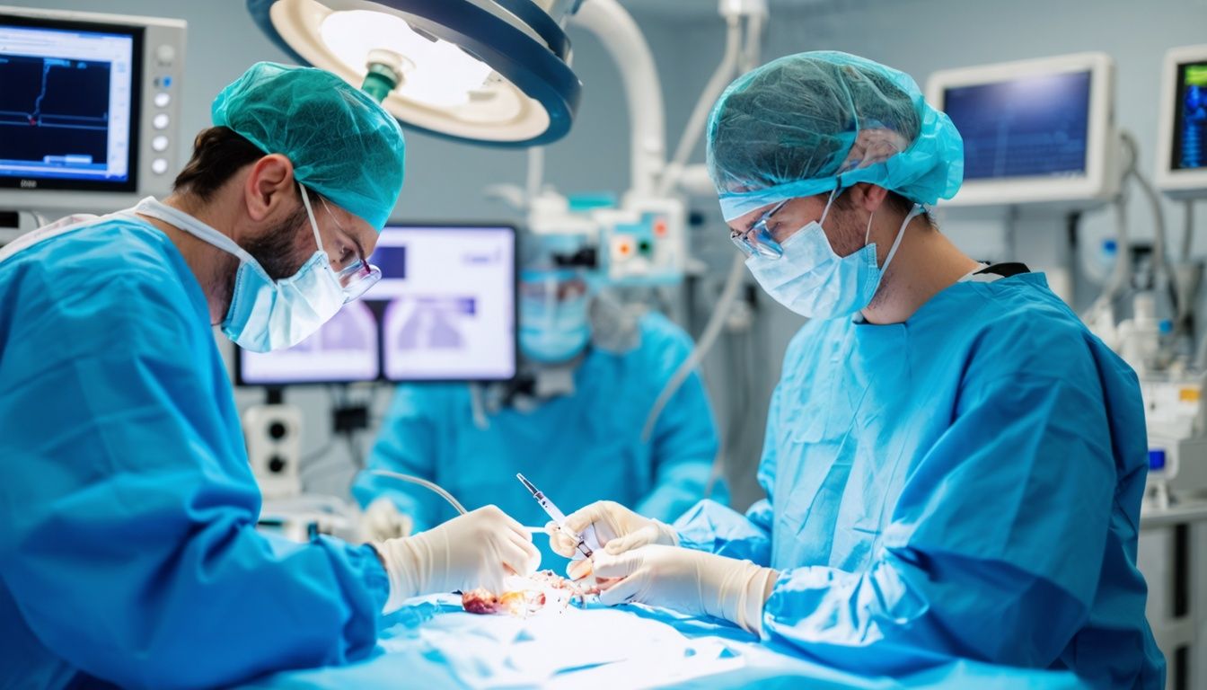
Endoscopic Submucosal Dissection (ESD) is a minimally invasive surgical technique used to remove precancerous and cancerous tumors in the gastrointestinal tract. It targets lesions under the lining of organs like the esophagus, stomach, and colon.
ESD allows doctors to precisely cut out tumors with safety margins, resulting in higher success rates and lower recurrence compared to other removal methods. 2
Preparing for ESD involves liquid diets, laxatives or enemas for bowel cleansing, and intravenous sedation during the one- to three-hour procedure. Key steps include marking tumor borders, injecting a solution to lift the tumor off the muscle wall, making a mucosal incision, and meticulously dissecting out the cancerous tissue using an electrosurgical knife – all guided by an endoscope camera.
Advanced techniques like high-pressure injections, traction systems, and carbon dioxide insufflation aid efficient tumor removal while reducing complications like bleeding or perforation. 1 Though highly effective for early gastrointestinal cancers, ESD requires specialized training limiting its widespread availability across the United States. Mastering this intricate procedure can lead to better outcomes for many patients. 3 The following sections explore ESD’s nuances and challenges in greater depth.
Key Takeaways
- Endoscopic submucosal dissection (ESD) is a minimally invasive procedure that allows complete removal of precancerous growths or early-stage cancers in the gastrointestinal tract, with a high success rate of over 99% and low recurrence risk of only 1%.
- The ESD procedure involves marking the lesion, submucosal injection, mucosal incision, and submucosal dissection, often aided by advanced traction systems and specialized tools like electrosurgical knives and coagulation devices.
- Proper preparation, including bowel cleansing and arranging transportation, is crucial for a safe and successful ESD procedure, and postoperative management with proton pump inhibitors, antacids, and dietary modifications supports recovery.
- Avoiding complications like bleeding and perforations during ESD requires meticulous technique, familiarity with hemostatic techniques and closure devices, and prompt recognition and management of adverse events.
- ESD offers advantages over other procedures, such as higher en-bloc and curative resection rates, preservation of organ structural integrity, and accurate pathological assessment, enabling expanded curative treatment for gastrointestinal neoplasms without invasive surgery.
Understanding Endoscopic Submucosal Dissection

Endoscopic submucosal dissection removes precancerous growths or early cancers from the gastrointestinal tract. It allows complete resection of lesions with a lower risk of complications compared to open surgery.
This minimally invasive procedure uses an endoscope inserted through the mouth or rectum. An electrosurgical knife dissects the lesion from the surrounding healthy tissue after injecting a solution beneath it.
What is Endoscopic Submucosal Dissection?
Endoscopic Submucosal Dissection (ESD) is a minimally invasive procedure. It removes precancerous and cancerous areas in the gastrointestinal (GI) tract. ESD targets tumors located under the lining of the GI tract.
It allows for complete removal with safety margins regardless of lesion size. 1
ESD is more effective than other methods like Endoscopic Mucosal Resection (EMR). It minimizes the risk of cancer spreading. I’ve performed countless ESDs on colorectal neoplasms and cancers of the esophagus.
The procedure involves marking the lesion, submucosal injection, mucosal incision, and submucosal dissection. Advanced traction systems aid efficient dissection. 2
As an experienced endoscopist, I find ESD invaluable. It facilitates “en bloc” resection (R0 resection) for large precancerous lesions. This precise technique prevents piecemeal removal.
It reduces the recurrence risk compared to EMR. ESD is transforming therapeutic endoscopy for GI neoplasms.
Who may benefit from this procedure?
Endoscopic submucosal dissection benefits patients with early-stage cancerous tumors or precancerous growths in the gastrointestinal tract. It provides an organ-preserving cure for early esophageal, gastric, and colorectal cancers that have not invaded deep layers. 3 Patients with Barrett’s esophagus and large colon polyps also qualify for this minimally invasive procedure.
The procedure removes tumors confined to the mucosal layer while preserving the underlying organ. It offers a high success rate of over 99% and low recurrence of only 1%. 2 ESD avoids major surgery and organ removal, decreasing complications and improving quality of life.
Early detection and appropriate treatment are crucial for managing gastrointestinal malignancies effectively.
Moving on to the next section, let’s explore the step-by-step procedure of endoscopic submucosal dissection.
Preparing for the Procedure
Preparing for an Endoscopic Submucosal Dissection involves specific steps. Patients follow a liquid diet and use laxatives or enemas to cleanse the bowel for a lower GI tract procedure. 4 Arranging for someone to drive them home after the one-to-three-hour procedure is essential. CO2 insufflation reduces pain and adverse events during the procedure. Careful preparation ensures a safe and successful Endoscopic Submucosal Dissection.
For the best outcomes, adherence to the prescribed bowel prep regimen is crucial. The colon must be thoroughly cleansed for optimal visibility during the endoscopy. Patients may experience some discomfort and frequent bowel movements as the laxatives take effect.
Proper hydration and following dietary instructions help minimize complications. With meticulous preparation, endoscopists can accurately mark lesions and proceed with the submucosal dissection technique. 3
The Procedure: Step by Step
ESD begins with marking the lesion. Doctors use special dyes to clearly identify margins. Next is submucosal injection. Doctors inject fluid under the lesion, lifting it off the muscle layer.
This makes it easier to remove the lesion. Doctors use specialized devices like the Flush knife. Injection solutions contain ingredients like hyaluronic acid. This makes the solution thick, aiding dissection by keeping the submucosal space expanded.
Marking the Lesion
The gastroenterologist initiates endoscopic submucosal dissection (ESD) by marking the lesion’s margins. Chromoendoscopy stains enhance visual delineation of the neoplastic area’s borders.
Accurate demarcation ensures complete resection without residual malignancy. 2Methylene blue or indigo carmine dye instillation helps define the circumferential extent before mucosal incision. The endoscopist may use coagulation markers, argon plasma coagulation (APC) or electrocautery to trace the planned perimeter.
Precise border marking facilitates subsequent submucosal dissection.
Lesion size, location and endoscopic appearance guide the marking technique chosen. Chromoendoscopy combined with virtual chromoendoscopy using narrow-band imaging (NBI) optimizes visualization during this critical step.
Careful outlining of the resection area is paramount for curative en bloc resection. 4
Submucosal Injection
After marking the lesion, the next crucial step is submucosal injection. This involves injecting a solution underneath the mucosal layer. Perpendicular injection into the mucosal plane aids in cutting deeper into the submucosa. 2 Viscous solutes injected at high pressure increase dissection speed by 25% compared to saline reinjection. 2 Solutions with limited diffusion and surface-active properties maintain a high-quality, durable lifting effect.
Hyaluronic acid, glycerol, and hydroxypropyl methylcellulose are examples of such solutions.
During my endoscopic submucosal dissection training, I learned the importance of proper submucosal injection technique. Injecting too superficially into the mucosal layer risks perforation.
Conversely, injecting too deep into the muscularis propria layer increases the risk of bleeding. 5 Finding the right plane and injecting the optimal viscous solution is crucial for safe and efficient dissection. 5 With practice, this technique becomes second nature, allowing for clean en bloc resection of lesions.
Mucosal Incision
A mucosal incision initiates the endoscopic submucosal dissection procedure. Electrosurgical knives create an incision around the lesion’s border. The incision extends through the mucosa, exposing the submucosal layer underneath.
Endocut I current facilitates a precise mucosal incision. The incision occurs at a distance from the target lesion, allowing access to the submucosal plane. 2 Trimming prevents flattening, maintaining favorable submucosal working space.
Appropriate mucosal incision lays the foundation for safe and efficient submucosal dissection. With experience, mastering the mucosal incision technique becomes essential for successful endoscopic submucosal dissections. 3
The next step involves submucosal injection to elevate the lesion.
Submucosal Dissection
Submucosal dissection commences after mucosal incision. The gastroenterologist uses an electrosurgical knife to dissect the submucosa beneath the lesion. Reinjection and injector knives help maintain a submucosal plane lift.
Traction devices like clips, rubber bands, and threads aid dissection speed and reduce perforation risks. CO2 insufflation is mandatory to reduce pain and adverse events. 6
During submucosal dissection, the lesion detaches from the muscularis propria layer. The endoscopist must meticulously dissect the submucosa away from the muscularis propria. Meticulous dissection prevents perforation and preserves en bloc resection.
Traction devices expose the submucosal plane, facilitating dissection and reducing procedure time. 4
Traction Systems
Traction systems enhance endoscopic submucosal dissection (ESD). Clip and line traction improves exposure and dissection speed. Double-clip traction also excels. Magnetic traction devices aid visualization.
Robotic endoscopes with traction capabilities exist. Elastic traction benefits small and large lesions alike. Novel low-cost traction techniques continuously develop.
Experiencing various traction approaches firsthand unveiled their merits. 7 Magnetic systems yielded unmatched views during a challenging gastric ESD case. Robotic assistance streamlined an esophageal procedure remarkably. 2 The next section examines avoiding complications.
Avoiding Complications
Managing bleeding during the procedure proves crucial. Physicians utilize advanced hemostatic techniques and tools to control any bleeding.
Perforations demand immediate attention. Endoscopic clipping devices help seal perforations. Surgeons may perform additional procedures if required.
Management of Bleeding
Bleeding control is paramount during endoscopic submucosal dissection (ESD). Techniques minimize intraoperative hemorrhage. Submucosal injection separates the mucosa from the muscle layer, reducing bleeding risk.
Understanding vascular architecture and high-density vessel areas is critical. Proper mucosal incision and dissection techniques prevent vessel injury. 8
Coagulation methods manage bleeding episodes effectively. Minor bleeding rarely causes significant issues. Serious hemorrhages require prompt hemostasis with hemoclips, coagulation forceps, or argon plasma coagulation.
Experience with these tools and quick decision-making are vital for safe ESDs. I’ve witnessed firsthand how meticulous bleeding control allows complete en bloc resections, optimizing cancer staging and treatment. 9
Management of Perforation
Controlling bleeding during an endoscopic submucosal dissection (ESD) procedure is crucial. However, preventing perforations and managing them effectively when they occur is equally important.
Perforations involve a hole or tear in the gastrointestinal tract wall. Endoscopic tissue closure devices like clips and suturing techniques help close these perforations. Complete closure is often achievable with these methods. 1 Experienced endoscopists should handle duodenal perforations as the risk is higher in this area.
Familiarity with closure devices is essential for endoscopists performing ESDs. Prompt recognition and appropriate management of perforations can prevent serious complications. Advanced tools and techniques enable safe and effective resections while minimizing adverse events. 10
Postoperative Management
After addressing bleeding and perforation risks during endoscopic submucosal dissection (ESD), postoperative care becomes crucial. Patients may experience a sore throat, upset stomach, vomiting, excessive gas, bloating, or cramping.
Monitoring for signs like blood in stools, chest pain, dizziness, fever, or vomiting blood is essential to detect complications promptly. 9
Proton pump inhibitors and antacids help reduce acid production and protect the dissected area. H2-receptor antagonists also aid in healing by blocking histamine receptors. Pain medication provides relief from discomfort.
Dietary modifications involving easily digestible foods and adequate hydration support recovery. Avoiding anti-coagulants minimizes bleeding risks. Endoscopic evaluation ensures proper healing before resuming regular activities. 2
Advancements and Considerations in ESD
Endoscopic submucosal dissection offers precise removal of cancers and precancerous lesions in the digestive tract. Novel techniques enhance visualization and control during the intricate procedure, enabling safer and more efficient dissections.
Read on to learn about the latest advancements.
Essential Prerequisite for Colonic ESD: Colonoscopy Without Loops
Performing a colonoscopy without loops is crucial for colonic endoscopic submucosal dissection (ESD). Looping occurs when the colonoscope forms an alpha or reverse alpha loop within the colon.
Looping prevents effective scope control and manipulation required for ESD. 2
Skilled endoscopists utilize techniques like shortening, underwater technique, and tube compression to minimize looping. These techniques facilitate straightening the colon, enabling smooth scope insertion and withdrawal.
Colonoscopy without loops ensures optimal visualization and access for safe, precise ESD dissection. 11
Mistakes to Avoid
Endoscopic submucosal dissection (ESD) is a challenging technique. Proper training and experience are crucial for success. 12
- Underestimating procedure duration. ESD procedures often take hours to complete. Rushing leads to complications. 3
- Inadequate submucosal injection. Insufficient submucosal elevation makes dissection difficult and risky.
- Incorrect incision line placement. Improper incision placement increases perforation and incomplete resection risks.
- Tenting the muscularis propria layer. Tenting this deepest layer raises perforation danger.
- Inadequate hemostasis. Overlooking minor bleeding points invites delayed bleeding.
- Forceful dissection. Excessive force can tear through layers, causing perforations.
- Continuing with poor visualization. Persistence without a clear view compromises safety.
- Inappropriate case selection. Attempting complex lesions before developing basic skills raises complication rates.
- Failure to recognize limitations. Attempting overly difficult cases exceeds one’s current capability.
- Lack of a dedicated team. Absence of experienced assistants and support staff hinders efficiency.
The next section discusses advancements and key considerations in ESD procedures.
Helpful Techniques for Efficient ESD
Mastering the Endoscopic Submucosal Dissection (ESD) technique requires diligent practice and familiarity with specific techniques. Efficient ESD procedures rely on proper preparation, skilled execution, and attention to detail.
- Utilize a high-definition endoscope with water-jet capabilities for optimal visualization and dissection.
- Employ a transparent cap attached to the endoscope tip to maintain a clear field of view during the procedure. 2
- Insufflate with carbon dioxide (CO2) gas, as it causes less pain and adverse events compared to air insufflation.
- Inject a viscous solution, such as hyaluronic acid or glycerol, into the submucosal layer for effective lifting and dissection.
- Use specialized ESD knives, such as the insulated-tip (IT) knife or the dual knife, for precise mucosal incisions and submucosal dissection.
- Implement traction techniques, like the clip-and-line method or the external grasping device, to counter the effects of gravity and improve visualization.
- Consider using magnetic anchor guidance systems for gastric lesions, enabling precise control and traction during dissection. 13
- Routinely perform endoscopic closure of mucosal defects with endoclips or over-the-scope clips to prevent adverse events.
- Engage in regular training and practice on animal models or simulators to enhance skills and proficiency in ESD techniques.
The Unique Challenges of Esophageal ESD
Esophageal endoscopic submucosal dissection presents unique obstacles. Surgeons confront limited maneuverability and visibility in the narrow esophageal lumen. The thin esophageal wall heightens perforation risks.
Submucosal fibrosis from esophageal squamous cell cancer or esophageal adenocarcinoma can impede safe dissection. Precise control over the electrosurgical knife is crucial to prevent injuring surrounding structures like the trachea or aorta.
Thorough preoperative staging utilizing endoscopic ultrasonography guides patient selection. Specialized techniques like the tunnel or double-scope methods aid esophageal ESD. Extensive training and meticulous endoscopic skills are prerequisites.
Esophageal perforations demand prompt management to avoid life-threatening complications. 3
Postoperative management poses distinct challenges. Esophageal strictures frequently occur post-ESD, necessitating periodic endoscopic balloon dilation. Bleeding risks persist due to esophageal vascular anatomy.
Close monitoring is mandatory. Oncologists advise adjuvant treatment strategies based on histopathological findings. Implementing quality control measures ensures optimal outcomes.
Potential advantages of esophageal ESD over conventional esophagectomy include organ preservation and faster recovery. However, appropriate patient selection based on multidisciplinary expertise remains paramount.
Comprehensive training, specialized instrumentation, and institutional experience underpin successful esophageal ESD programs. Discussing essential prerequisites and training algorithms will elucidate this complex technique further. 12
Advantages of ESD over Other Procedures
ESD yields higher en-bloc and curative resection rates compared to endoscopic mucosal resection (EMR). It enables complete removal of previously unresectable lesions like large colorectal adenomas or early gastric cancers. 3 ESD preserves the structural integrity of the gastrointestinal tract. It allows precise pathological assessment by providing en-bloc resected tissue samples. 14
I have first-hand experience assisting gastroenterologists during ESD procedures. The higher en-bloc resection rate with ESD minimizes recurrence risk. It facilitates accurate histological assessment of lateral and deep margins.
ESD permits expanded curative treatment for gastrointestinal neoplasms without invasive surgery. Patients benefit from quicker recovery times compared to conventional surgical resection modalities.
Conclusion
Endoscopic submucosal dissection demands precision and expertise. This cutting-edge technique empowers clinicians to remove precancerous lesions and early-stage cancers from the gastrointestinal tract.
Its minimally invasive nature enhances patient recovery, minimizing complications. Mastering ESD unlocks new frontiers in personalized cancer care, staging tumors accurately. While challenging, centers specializing in this procedure are paving the way for improved outcomes.
The future of gastrointestinal oncology lies in such advanced, targeted therapies.
FAQs
1. What is Endoscopic Submucosal Dissection (ESD)?
ESD is a minimally invasive procedure. It removes early-stage cancers from the digestive tract. Doctors use it for esophageal, stomach, and colorectal cancers.
2. How does ESD differ from other treatments?
ESD is more precise than endoscopic mucosal resections. It allows for complete removal of larger lesions. This technique avoids major surgery like removing part of the esophagus.
3. What conditions can ESD treat?
ESD treats early-stage cancers and precancerous lesions. It’s used for esophageal squamous cell carcinoma, gastric carcinoma, and colorectal polyps.
4. Is ESD better than traditional surgery?
Yes, for certain cases. ESD is less invasive than laparoscopic surgery. It preserves organ function and reduces recovery time. It’s effective for lesions without lymph node metastasis.
5. How do doctors determine if ESD is suitable?
Doctors use endoscopy and biopsy. They check the depth of tumor invasion. They look at the lamina propria and lymphatic involvement. Histological examination helps decide treatment.
6. What are the risks of ESD?
Risks include bleeding and perforation. Rectal bleeding can occur after colorectal ESD. Gastric ulcers may form after stomach procedures. Proper training reduces these risks.
References
- ^ https://www.cghjournal.org/article/S1542-3565(18)30807-3/fulltext
- ^ https://www.ncbi.nlm.nih.gov/pmc/articles/PMC8589544/
- ^ https://www.ncbi.nlm.nih.gov/pmc/articles/PMC3742702/
- ^ https://www.hopkinsmedicine.org/health/treatment-tests-and-therapies/endoscopic-submucosal-dissection
- ^ https://www.giejournal.org/article/S0016-5107(21)00087-0/pdf
- ^ https://my.clevelandclinic.org/health/treatments/21091-endoscopic-submucosal-dissection-esd-for-esophageal-and-gastric-cancer
- ^ https://www.researchgate.net/publication/365023301_Advancing_endoscopic_traction_techniques_in_endoscopic_submucosal_dissection
- ^ https://www.ncbi.nlm.nih.gov/pmc/articles/PMC7008155/
- ^ https://www.ncbi.nlm.nih.gov/pmc/articles/PMC3180012/
- ^ https://www.ncbi.nlm.nih.gov/pmc/articles/PMC6453857/
- ^ https://www.ncbi.nlm.nih.gov/pmc/articles/PMC8349195/
- ^ https://www.ncbi.nlm.nih.gov/pmc/articles/PMC10253693/
- ^ https://pubmed.ncbi.nlm.nih.gov/34790536/
- ^ https://www.ncbi.nlm.nih.gov/pmc/articles/PMC4948268/