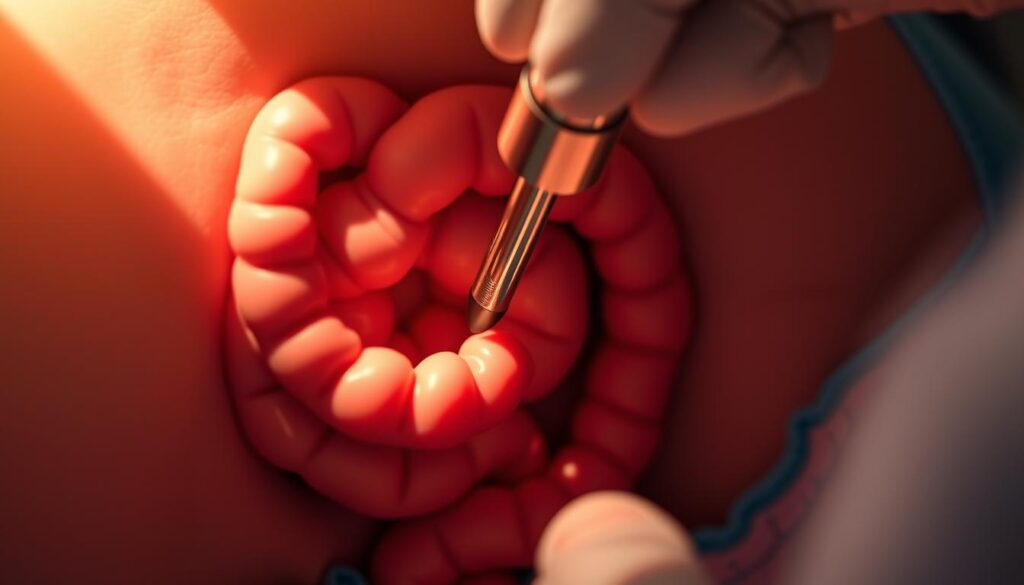
Crohn’s disease, a form of inflammatory bowel disease, requires precise diagnostic and monitoring tools for effective management. Among these, endoscopy plays a pivotal role. It allows clinicians to visualize the gastrointestinal tract, assess disease severity, and guide treatment decisions.
Modern advancements in endoscopic technology have expanded its applications, enabling more accurate evaluations. Standardized scoring systems provide objective measures of disease activity, while mucosal healing has emerged as a critical treatment endpoint. These developments underscore the importance of endoscopy in contemporary care paradigms.
Key Takeaways
- Endoscopy is essential for diagnosing and monitoring Crohn’s disease.
- It informs therapeutic decisions by providing detailed visual insights.
- Advanced technologies enhance the accuracy of endoscopic evaluations.
- Standardized scoring systems offer objective disease assessment.
- Mucosal healing is a key treatment goal in modern management strategies.
Introduction to Endoscopy in Crohn’s Disease
Flexible imaging technology plays a critical role in diagnosing gastrointestinal disorders. These procedures, known as endoscopy, utilize high-definition cameras to provide detailed views of the mucosal lining. This approach is particularly valuable in managing inflammatory bowel conditions, where precise visualization is essential.
What is Endoscopy?
Endoscopy involves the use of a flexible tube equipped with a camera to examine the digestive tract. It allows clinicians to visualize areas from the esophagus to the terminal ileum. This method is crucial for obtaining targeted biopsies, which provide histologic specimens for accurate diagnosis.
Why is Endoscopy Important in Crohn Disease?
In Crohn disease, endoscopic findings help differentiate it from other bowel disease types, such as ulcerative colitis. Characteristic features like skip lesions and transmural inflammation are identifiable through this technique. Additionally, endoscopy is indispensable for monitoring disease progression and treatment response over time.
Diagnostic Role of Endoscopy in Crohn’s Disease
Visualizing the digestive tract is essential for identifying inflammatory disorders. This process relies on advanced imaging techniques, such as colonoscopy and upper gastrointestinal endoscopy. These methods provide detailed insights into mucosal changes, enabling accurate diagnosis and effective management.
Colonoscopy and Flexible Sigmoidoscopy
Colonoscopy is a cornerstone in evaluating the ileocolonic region. It involves intubation of the terminal ileum, which is critical for initial assessments. Site-specific biopsy collection during this procedure helps confirm diagnosis and differentiate between conditions like Crohn’s disease and ulcerative colitis. Skip lesions and aphthous ulcers are key endoscopic features that aid in this differentiation.
Flexible sigmoidoscopy, while useful, has limitations in severe colitis cases. It provides a partial view of the colon, making it less comprehensive than a full colonoscopy. However, it remains a valuable tool for targeted evaluations in specific clinical scenarios.
Upper Gastrointestinal Endoscopy
Upper gastrointestinal endoscopy (EGD) is increasingly recognized for its role in detecting gastric involvement. Studies show that 93.5% of H. pylori-negative patients exhibit chronic gastritis. Bamboo joint-like gastric lesions, pathognomonic for Crohn’s disease, are often identified through this method.
Pediatric populations, in particular, benefit from EGD due to the growing awareness of upper GI involvement in younger patients. This technique complements ileocolonic examinations, providing a holistic view of disease activity and guiding tailored treatment plans.
Advanced Endoscopic Technologies
Innovative technologies are transforming the way clinicians assess and manage digestive disorders. These advancements provide detailed insights into the bowel, enabling precise diagnosis and tailored treatment plans. Among these, wireless capsule endoscopy and balloon-assisted enteroscopy stand out as game-changers.
Capsule Endoscopy
Wireless capsule endoscopy offers a non-invasive method for evaluating the small bowel. With a diagnostic yield of 71%, it is particularly effective in identifying lesions and abnormalities. The PillCam SB3, equipped with adaptive frame rate technology, ensures high-quality imaging while minimizing symptoms of discomfort for patients.
However, the risk of capsule retention necessitates prior patency assessment. This ensures safety and prevents complications during the procedure.
Balloon-Assisted Enteroscopy
Balloon-assisted enteroscopy plays a critical role in therapeutic interventions. It allows clinicians to access deep segments of the gastrointestinal tract, enabling biopsies and treatments. This method is particularly valuable for patients with complex conditions requiring precise management.
Compared to radiologic modalities, balloon-assisted enteroscopy offers superior diagnostic accuracy. Safety protocols in specialized centers further enhance its reliability and effectiveness.
- Wireless capsule endoscopy provides non-invasive imaging of the small bowel.
- Balloon-assisted enteroscopy enables deep therapeutic interventions.
- Both technologies enhance diagnostic accuracy and patient care.
Endoscopic Scoring Systems
Accurate assessment of gastrointestinal conditions relies on standardized scoring systems. These tools provide a structured approach to evaluating disease severity and guiding treatment decisions. By offering objective metrics, they enhance the precision of clinical evaluations and improve patient care.
Mayo Endoscopic Subscore
The Mayo Endoscopic Subscore is a four-grade scale that assesses mucosal changes, ranging from erythema to severe ulceration. This system is widely used in clinical trials and routine practice to evaluate disease activity in the colon. Its simplicity and reproducibility make it a valuable tool for consistent monitoring.
Simple Endoscopic Score for Crohn’s Disease (SES-CD)
The SES-CD provides a quantitative evaluation of disease severity across five intestinal segments. It assesses ulcer size, affected surface area, and narrowing, offering a comprehensive view of disease activity. International consensus recommends reassessment every 6-9 months to monitor treatment response and disease progression.
Standardized scoring systems like the SES-CD and Mayo Subscore benefit multicenter trials by ensuring consistent data comparison. They also correlate with long-term outcomes, highlighting their importance in management strategies. Achieving mucosal healing, a key treatment goal, is often measured using these metrics.
- The Mayo Subscore evaluates mucosal changes from erythema to ulceration.
- The SES-CD quantifies disease severity across multiple intestinal segments.
- Standardization enhances data comparison in clinical trials.
- Scoring systems correlate with long-term patients outcomes.
Endoscopy in Disease Management
Modern medical strategies emphasize the importance of accurate disease assessment. Endoscopic techniques play a critical role in evaluating the gut and guiding therapeutic decisions. These methods provide detailed visual insights, enabling clinicians to monitor disease activity and optimize management plans.
Assessing Disease Activity
Endoscopic evaluations are essential for determining the severity of gastrointestinal conditions. High-definition imaging allows for the detection of subtle inflammatory changes, which are often missed in standard assessments. This precision is crucial for tailoring treatment strategies to individual patients.
For example, mucosal healing, confirmed through colonoscopy, is associated with a 52% reduction in surgery rates over five years. This highlights the long-term benefits of achieving endoscopic remission in clinical practice.
Monitoring Treatment Response
Regular endoscopic surveillance is key to evaluating the effectiveness of biologic therapies. Protocolized intervals ensure that treatment plans are adjusted based on objective findings. This approach aligns with the SELECT initiative’s treat-to-target recommendations, which emphasize tight control strategies.
Comparing histologic and endoscopic remission concepts further enhances clinical decision-making. While histologic remission focuses on microscopic healing, endoscopic remission provides a broader view of mucosal recovery.
- Endoscopic confirmation of biologic therapy effectiveness improves outcomes.
- Protocolized surveillance intervals optimize management plans.
- High-definition imaging detects subtle inflammatory changes in the gut.
- Tight control strategies reduce hospitalization rates for patients.
| Key Aspect | Role in Management |
|---|---|
| Mucosal Healing | Reduces surgery rates by 52% over five years |
| HD Imaging | Detects subtle inflammatory changes |
| Protocolized Surveillance | Optimizes treatment adjustments |
| Tight Control Strategies | Lowers hospitalization rates |
Endoscopic Therapy in Crohn’s Disease
Managing complex gastrointestinal disorders often involves targeted endoscopic procedures. These techniques are essential for addressing complications like strictures and fistulae, which are common in chronic bowel conditions. By providing precise interventions, endoscopic therapy improves outcomes and enhances quality of life for patients.

Stricture Dilation
Endoscopic balloon dilation (EBD) is a primary method for treating strictures, with an 80% success rate at one year. The procedure involves expanding narrowed segments using a balloon catheter, typically up to 20mm in diameter. This approach alleviates symptoms such as abdominal pain and obstruction.
However, the risk of perforation remains at 3%, necessitating careful patient selection. Emerging techniques like needle-knife stricturotomy and self-expanding metal stent (SEMS) placement offer additional options for refractory cases. Post-procedural monitoring is critical to detect complications early and ensure optimal recovery.
Fistula Management
Fistulizing disease presents unique challenges, often requiring multidisciplinary care. Endoscopic vacuum therapy has shown promise in managing complex fistulae, promoting healing and reducing infection risks. Contraindications for EBD in these cases include active sepsis or extensive tissue damage.
Regular follow-up and imaging are essential to evaluate treatment response. Surgical referral criteria are based on factors like fistula location, severity, and patient history. This collaborative approach ensures comprehensive care tailored to individual needs.
- Endoscopic balloon dilation achieves 80% success in stricture treatment.
- Needle-knife stricturotomy and SEMS placement are emerging alternatives.
- Endoscopic vacuum therapy is effective for complex fistulae.
- Post-procedural monitoring detects complications early.
- Multidisciplinary decision-making optimizes treatment plans.
Post-Surgical Endoscopic Evaluation
Post-surgical evaluation of the gastrointestinal tract is critical for assessing treatment outcomes and detecting complications. This process involves detailed examination of reconstructed anatomy, such as the ileal pouch and neoterminal ileum. These evaluations help identify issues like strictures, inflammation, or recurrence of disease.
Ileal Pouch-Anal Anastomosis (IPAA)
IPAA is a common surgical procedure for patients with ulcerative colitis. Pouchoscopy, often performed using gastroscopes, allows clinicians to assess the anastomotic site. The Rutgeerts score is a valuable tool for predicting surgical recurrence risk based on endoscopic findings.
Key features to evaluate include pouchitis versus Crohn’s pouch disease. Pouchitis typically presents with erythema and ulceration, while Crohn’s pouch disease may show skip lesions. Histologic sampling during pouchoscopy aids in accurate diagnosis and guides further management.
Neoterminal Ileum Examination
Examination of the neoterminal ileum is essential for detecting postoperative complications. Surveillance intervals are recommended every 6-12 months, depending on disease activity. Balloon dilation techniques are often employed to manage anastomotic strictures, improving patients outcomes.
Differential diagnosis of cuffitis, a condition affecting the rectal cuff, is also critical. Endoscopic features include erythema and friability, which must be distinguished from other inflammatory conditions. Histologic evaluation provides additional insights into the underlying pathology.
- Pouchoscopy protocols assess anastomotic integrity and detect complications.
- The Rutgeerts score predicts recurrence risk in IPAA patients.
- Balloon dilation techniques are effective for managing strictures.
- Histologic sampling ensures accurate diagnosis of cuffitis and pouchitis.
- Regular surveillance of the neoterminal ileum is recommended for optimal care.
Endoscopy in Pediatric Crohn’s Disease
Pediatric cases of gastrointestinal disorders require specialized diagnostic approaches. Unlike adults, children present unique challenges that demand tailored protocols and careful consideration of growth parameters. These factors are critical for accurate diagnosis and effective management.
Unique Considerations in Children
Age-specific sedation protocols and scope selection are essential in pediatric care. Smaller, flexible scopes are often used to minimize discomfort and ensure safety. Additionally, integrating growth parameters with endoscopic findings provides a comprehensive view of the child’s health.
Differentiating Crohn’s disease from eosinophilic gastrointestinal disorders can be challenging. Endoscopic features like skip lesions and mucosal inflammation must be carefully evaluated. Early detection and accurate diagnosis are crucial for long-term outcomes.
Upper GI Involvement in Pediatric Patients
Studies show that 50% of pediatric cases involve upper gastrointestinal regions. Esophageal and gastric disease is more prevalent in children compared to adults. This highlights the importance of upper GI endoscopy, even in the absence of symptoms.
Early upper GI involvement can have long-term implications. Regular monitoring and timely intervention are necessary to prevent complications like infection and severe inflammation. Collaborative care between gastroenterologists and pediatricians ensures optimal outcomes for young patients.
Endoscopy and Colorectal Cancer Surveillance
Colorectal cancer surveillance is a critical component in managing inflammatory bowel conditions. Patients with chronic colitis, particularly ulcerative colitis, face a 13-fold increased risk of developing colorectal cancer compared to the general population. Advanced endoscopic techniques, such as chromoendoscopy, play a pivotal role in early dysplasia detection and effective management.
Chromoendoscopy and Dysplasia Detection
The SCENIC consensus emphasizes the superiority of chromoendoscopy over random biopsies for dysplasia detection. Targeted biopsies using methylene blue or indigo carmine dye application significantly improve diagnostic yield. This technique enhances the visualization of subtle mucosal changes, enabling precise identification of precancerous lesions.
Management of Dysplastic Lesions
Endoscopic mucosal resection (EMR) is the preferred method for removing visible dysplastic lesions. For patients with primary sclerosing cholangitis and colitis, surveillance intervals are recommended every 6-12 months. Pathologic criteria differentiate indefinite dysplasia from confirmed cases, guiding further management.
- Chromoendoscopy increases dysplasia detection accuracy compared to random biopsies.
- Methylene blue and indigo carmine enhance mucosal visualization.
- EMR techniques are effective for removing visible lesions.
- Surveillance intervals are tailored based on individual risk factors.
- Pathologic evaluation ensures accurate diagnosis and treatment planning.
Complications and Risks of Endoscopy
Every medical procedure carries inherent risks that require careful management. While endoscopy is generally safe, understanding potential complications is essential for ensuring patient safety and optimal outcomes. Clinicians must weigh these risks against the benefits of the procedure, especially in complex cases.

Infection and Perforation Risks
Infection is a rare but serious complication, particularly in immunocompromised patients. Antibiotic prophylaxis is often recommended to mitigate this risk. Perforation, though uncommon, occurs in 0.1-0.3% of diagnostic colonoscopies. Severe colitis is a relative contraindication due to the heightened risk of perforation.
Endoscopic clipping is a preferred method for managing small perforations, while surgical intervention may be necessary for larger ones. Post-polypectomy syndrome, characterized by fever and abdominal pain, requires prompt recognition and management to prevent further complications.
Managing Adverse Events
Risk stratification models help identify patients at higher risk for procedure-related complications. These models consider factors like age, medical history, and the condition of the body. For pregnant individuals, radiation-free alternatives are recommended to minimize risks to both the mother and fetus.
Adverse events, such as bleeding or allergic reactions, require immediate attention. Clinicians must be prepared with emergency protocols and ensure that all equipment is readily available. Regular training and adherence to safety guidelines are critical for minimizing risks and ensuring patient safety.
- Antibiotic prophylaxis reduces infection risks in immunocompromised patients.
- Endoscopic clipping is effective for small perforations.
- Risk stratification models identify high-risk individuals.
- Radiation-free alternatives are safer for pregnant patients.
- Emergency protocols ensure prompt management of adverse events.
Patient Preparation for Endoscopy
Proper preparation is critical for ensuring the success of diagnostic evaluations. It involves dietary restrictions and bowel cleansing to optimize visualization during the procedure. These steps are essential for accurate results and minimizing risks.
Dietary Restrictions
A clear liquid diet is standard 24 hours before the procedure. This includes water, broth, and gelatin, which help clear the digestive tract. For patients with strictures, a low-residue diet may be recommended to reduce complications.
Pediatric-specific protocols often involve age-appropriate dietary adjustments. These modifications ensure safety and comfort for younger patients while maintaining preparation effectiveness.
Bowel Preparation
Split-dose polyethylene glycol (PEG) regimens are widely used for optimal bowel cleansing. This method involves taking half the dose the evening before and the remainder on the morning of the procedure. It enhances visualization quality and reduces the need for repeat tests.
Electrolyte disturbances during preparation require careful management. Clinicians may provide support through hydration strategies or electrolyte supplements to maintain balance.
- Clear liquid diets ensure a clean digestive tract.
- Split-dose PEG regimens improve cleansing effectiveness.
- Pediatric protocols address unique preparation needs.
- Electrolyte management prevents complications.
- Patient education enhances compliance and outcomes.
| Preparation Step | Purpose |
|---|---|
| Clear Liquid Diet | Clears the digestive tract for better visualization |
| Split-Dose PEG | Optimizes bowel cleansing effectiveness |
| Low-Residue Diet | Reduces complications in stricturing patients |
| Pediatric Protocols | Ensures safety and comfort for younger patients |
| Electrolyte Management | Prevents imbalances during preparation |
Future Directions in Endoscopic Technology
The evolution of diagnostic tools is reshaping the landscape of gastrointestinal healthcare. Advanced technologies are enhancing the precision and efficiency of endoscopic procedures, offering new opportunities for improved patient care. These innovations are driven by a growing interest in integrating cutting-edge solutions into clinical practice.
Innovations in Endoscopic Imaging
Confocal laser endomicroscopy is emerging as a groundbreaking tool for real-time histologic prediction. This technique provides detailed information at the cellular level, enabling clinicians to make immediate diagnostic decisions. Another advancement is molecular imaging, which uses fluorescent probes to highlight specific biomarkers in tissue.
Robotic-assisted platforms are also gaining traction, offering enhanced precision and control during procedures. These systems reduce operator fatigue and improve outcomes, particularly in complex cases. Telemedicine applications are further expanding the reach of endoscopic monitoring, allowing remote consultations and follow-ups.
Role of Artificial Intelligence in Endoscopy
Artificial intelligence (AI) is playing a transformative role in endoscopic evaluations. AI algorithms, such as those in Rapid Reader® software, automate scoring systems like SES-CD, reducing human error and increasing efficiency. These tools analyze vast datasets to identify patterns and predict disease progression.
Molecular profiling of the gastric microbiome is another area where AI is making strides. By analyzing microbial data, AI can provide insights into disease mechanisms and potential treatment targets. This integration of AI into endoscopy is paving the way for more personalized and effective healthcare solutions.
- Confocal laser endomicroscopy enables real-time histologic predictions.
- AI algorithms automate SES-CD scoring for objective assessments.
- Molecular imaging with fluorescent probes enhances biomarker detection.
- Robotic-assisted platforms improve precision in complex procedures.
- Telemedicine expands access to endoscopic monitoring and consultations.
Conclusion
The integration of advanced imaging techniques has revolutionized gastrointestinal care. Endoscopy remains central to the management and diagnosis of Crohn disease, providing critical insights into disease activity and treatment response. Technological advancements, such as high-definition imaging and AI algorithms, have significantly improved diagnostic accuracy and efficiency.
Standardized protocols ensure consistency across care settings, enhancing patient outcomes. The anticipated integration of AI into therapeutic decision-making promises to further refine treatment strategies. Continued research into minimally invasive techniques will drive innovation, offering safer and more effective options for patients.