Key Takeaways
- Clinical Bottom Line
- What Changed
- Mapping Biopsies
- Surveillance Decisions
Clinical Bottom Line
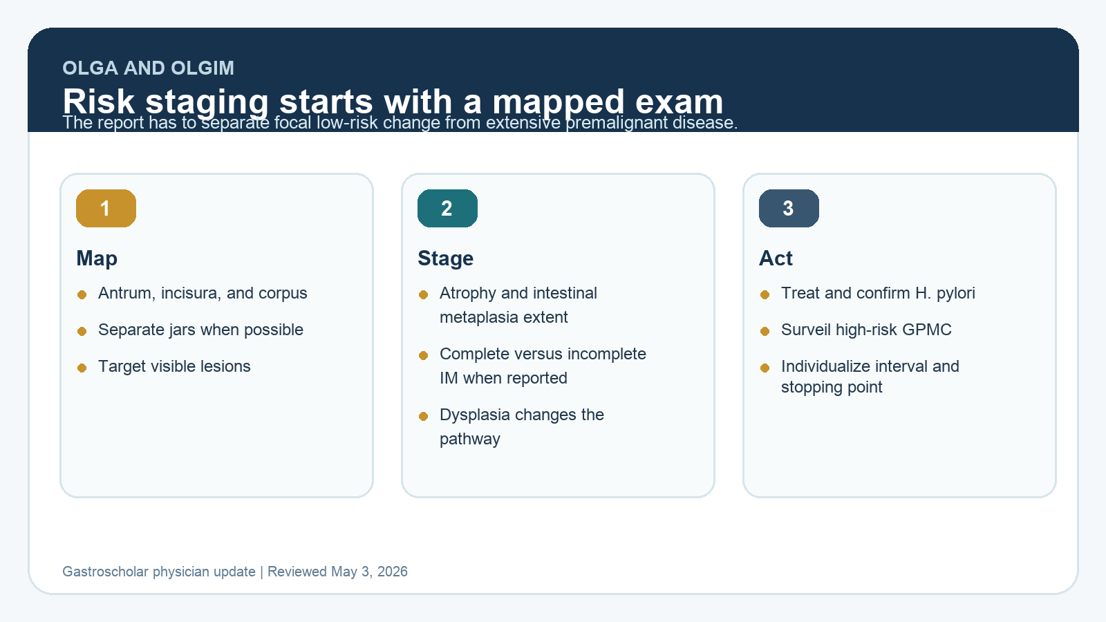
OLGA and OLGIM staging only help if the endoscopy and biopsy workflow is built to support them. The practical task is to identify visible lesions, map the stomach when staging is needed, separate antrum/incisura from corpus specimens, and use the final histology to decide whether the patient has limited low-risk change or extensive gastric premalignant disease that deserves surveillance.
| Element | What It Means | Practice Implication |
|---|---|---|
| OLGA | Stages gastric atrophy based on severity and extent. | Useful when gland loss is the dominant pathology question. |
| OLGIM | Stages gastric intestinal metaplasia based on severity and extent. | Often more reproducible than atrophy grading and clinically intuitive for surveillance decisions. |
| Sydney-style mapping | Samples antrum, incisura, and corpus in a structured way. | Prevents a single bottle of gastric biopsies from becoming an uninterpretable risk signal. |
| Dysplasia | Changes management from routine surveillance to expert review and possible resection or short-interval reassessment. | Visible lesions should be targeted and described, not blended into mapping biopsies. |
What Changed
The 2025 ACG guideline on gastric premalignant conditions pushes U.S. practice toward a more organized stomach surveillance model. The stomach is starting to look more like the colon and Barrett’s esophagus in one important way: risk stratification depends on quality baseline staging. A pathology phrase like “intestinal metaplasia present” is not enough to make a durable surveillance decision.
Mapping Biopsies
When gastric atrophy or gastric intestinal metaplasia needs staging, the endoscopist should perform a careful visual exam first, then obtain mapping biopsies. Visible abnormalities should be targeted and submitted separately. Mapping biopsies should be labeled by location, commonly separating antrum/incisura and corpus specimens. This allows the pathologist to assess extent and helps the clinician distinguish focal antral GIM from extensive disease involving the corpus.
OLGA and OLGIM are not magic scores. They are structured ways to convert histology and anatomic extent into risk categories. Their value falls apart if the biopsy sites are unknown or if all tissue is placed into one jar. The report should tell the next clinician where the biopsies came from and why they were taken.
Surveillance Decisions
Surveillance is most relevant for patients at higher risk of progression, including extensive atrophy or intestinal metaplasia, incomplete-type intestinal metaplasia when reported, family history of gastric cancer, persistent H. pylori, autoimmune gastritis, dysplasia history, or other high-risk context. Many patients with limited, low-risk findings will not need the same intensity of follow-up.
A common practical interval for higher-risk gastric premalignant conditions is around 3 years, but the interval should not be automatic. It should be individualized by quality of the baseline exam, pathology, patient risk, comorbidity, age, preferences, and whether H. pylori was treated and eradication confirmed.
How To Apply This In Practice
- Do not bury visible lesions in mapping biopsies. Target and label them separately.
- Ask pathology partners to report extent, incomplete versus complete intestinal metaplasia when feasible, dysplasia, and autoimmune gastritis features.
- Confirm H. pylori eradication in every patient with gastric premalignant conditions.
- Use OLGA or OLGIM language when it helps the next clinician understand risk.
- Document why surveillance is recommended, deferred, or stopped.
Selected Sources
- PubMed: ACG Clinical Guideline on Gastric Premalignant Conditions
- ACG gastric premalignant conditions guideline highlights
- ESGE MAPS III guideline update
Clinical update for gastroenterologists and endoscopists. Reviewed May 2026.