GI Endoscopy · 8 min read
Sessile Serrated Lesions in 2026: Mucus Cap, WASP, Complete Resection
Pale, flat, right-sided, often under a mucus cap. NICE 1 does not separate SSL from HP. WASP does. Take it out completely. Count SSLDR.
WHO 2019 renamed SSA/P to sessile serrated lesion. One unequivocal distorted crypt is enough. These are the pale, mucus-capped, right-colon lesions that drive a large share of post-colonoscopy cancer. ADR does not count them. SSLDR of at least 6% does. Recognition without complete resection is how CARE turned into interval cancer.
Experienced teaching points
Clinical Pearls
- Wash the cap. The lesion is the pale cloud under the mucus, not the mucus.
- NICE 1 is HP or SSL. WASP adds cloud-like surface, indistinct border, irregular shape, and dark spots in crypts. Two of those four after NICE 1 is SSL.
- Anything pale and mucus-capped proximal to the sigmoid is SSL until pathology says otherwise. Retrieve it. Do not suck it away.
- CARE left residual neoplastic tissue in 31% of sessile serrated lesions versus 7% of conventional adenomas. Cold forceps is not a resection.
- SSLDR minimum is 6% on the same denominator idea as ADR. Dashboard: the colonoscopy quality page.
Clinical Bottom Line
| Question | 2026 practical answer |
|---|---|
| What do I call it? | Sessile serrated lesion (SSL). WHO 2019 dropped SSA/P. Traditional serrated adenoma is a different polyp. Hyperplastic polyp is the rectosigmoid diminutive cousin, not the proximal mucus cap. |
| How do I see it? | Right colon, pale, flat or sessile, cloud-like, indistinct edge, often a mucus cap or a rim of debris. Wash. Then look again. |
| Optical diagnosis | NICE 1 does not split HP from SSL. WASP (Hazewinkel features): after NICE 1, two of cloud-like surface, indistinct border, irregular shape, dark spots inside crypts. |
| Pathology threshold | One unequivocal architecturally distorted crypt (dilated, boot- or L-shaped base, growth along muscularis mucosae) is enough in WHO 2019. Do not wait for three crypts. |
| Take it out how? | Cold snare diminutive and small. EMR or UEMR for 10 to 20 mm sessile. Wide enough to bury the edge. Piecemeal large SSL: tattoo and look at 6 months. |
| SSLDR | At least 6% on screening, surveillance, and diagnostic exams from age 45, same exclusions as ADR. Details: colonoscopy quality page. |
| Surveillance | USMSTF 2020: 1 to 2 SSL under 10 mm, 5 to 10 years. Three or four, 3 to 5. Five to ten, any SSL 10 mm or larger, or SSL with dysplasia: 3 years. Table: surveillance page. |
Why did the name change from SSA/P to SSL?
WHO 5th edition (2019) uses sessile serrated lesion. The older names (sessile serrated adenoma, sessile serrated polyp, SSA/P) described the same thing and argued about whether a lesion without cytologic dysplasia deserved the word adenoma. It does as a cancer precursor. It does not as a NICE 2 brown-vessel adenoma. SSL is the compromise. Use it. Pathology reports that still say SSA/P are the same lesion.
Traditional serrated adenoma is not an SSL. TSA is usually distal, protuberant, eosinophilic, with ectopic crypts. Different pathway, same 3-year USMSTF interval once you have one.
Hyperplastic polyps are still real. Diminutive, pale, NICE 1, rectosigmoid. They do not count toward SSLDR. A pale mucus-capped lesion in the ascending colon is not "just a hyperplastic." Retrieve it.
How do I recognize one on withdrawal?
The lesion hides as a fold, a mucus puddle, or "normal" pale mucosa. Tadepalli's morphologic series and Hazewinkel's expert stills agree on the same tells:
- Proximal to the splenic flexure more often than not.
- Flat or sessile, not a mushroom.
- Color close to surrounding mucosa. You are looking for a cloud, not a cherry.
- Mucus cap, or a rim of bubbles and debris that outlines an otherwise invisible edge.
- Indistinct border. If you can draw a sharp line around it on white light, you may be looking at an HP or an adenoma.

Hazewinkel 2013 (PMID 23433877): on white light, indistinct borders and a cloud-like surface independently predicted SSL. On NBI, add irregular shape and dark spots inside the crypts (open, dilated pits). IJspeert's WASP classification (PMID 25753029) turned that into a sequence: start with NICE. If NICE 2 or 3, you are in adenoma/cancer land. If NICE 1, score the four Hazewinkel features. Two or more: call it SSL. Fewer: call it HP.
NICE 1 is necessary and not sufficient. A validated rectosigmoid resect-and-discard program for diminutive NICE 1 polyps is a different argument. It does not apply to the right colon, and it does not apply to a 12 mm mucus cap anywhere.
What does pathology need to see?
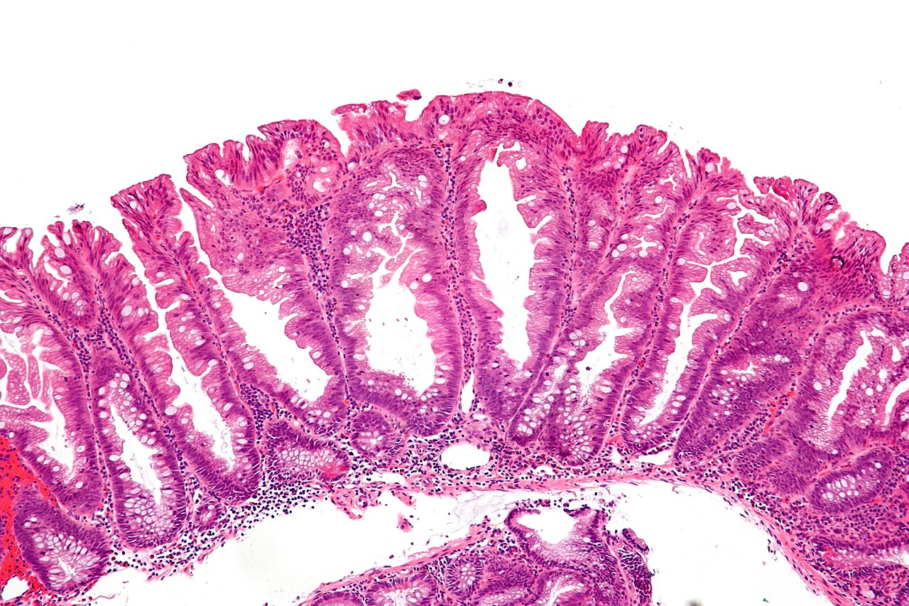
WHO 2019: one unequivocal architecturally distorted serrated crypt. Distortion means basal dilation, serration that reaches the base, a boot or L, or a crypt that runs horizontal along the muscularis mucosae. Older rules that demanded two or three such crypts under-called the lesion. If your group SSLDR is stuck under 6% and the endoscopists are finding mucus caps, talk to pathology before you buy a cap attachment.
Cytologic dysplasia inside an SSL is a different lesion (SSL-D). Treat it as an advanced polyp: complete resection, 3-year interval, and do not leave the dysplastic nodule because the rest looks like a fold. Endoscopically the dysplastic focus often looks like a conventional adenoma sitting on a pale carpet.
BRAF plus CIMP is the molecular story. It explains why some of these move faster than the classic 10-year adenoma sequence, especially on the right. It does not change tonight's resection.
How do I get it all out?
Rex 2012: remove every serrated lesion proximal to the sigmoid, and every rectosigmoid serrated lesion larger than 5 mm. That recommendation has not aged out.
Pohl's CARE study (PMID 23022496) is why "I took a piece" is not a plan. Incomplete resection was 10% overall for 5 to 20 mm nonpedunculated neoplasia, 17% at 10 to 20 mm, and 31% for sessile serrated lesions versus 7% for conventional adenomas. The residual sat at the edge. Cold forceps for a 6 mm SSL is how you manufacture a 2029 right-colon cancer.
- Diminutive and small: cold snare, including a rim of normal. Retrieve. Do not suction-and-hope in the right colon.
- 10 to 20 mm sessile: UEMR or conventional EMR. Underwater helps the pale edge lift into view.
- Larger: piecemeal EMR is acceptable. Tattoo. Look in 3 to 6 months. USMSTF puts piecemeal lesions 20 mm or larger at 6 months.
If the pathologist says SSL and you thought you left a clean snare line, believe CARE more than your memory of the edge.
What interval do I assign?
USMSTF 2020 (Gupta, PMID 32044092), after a high-quality exam:
- 1 to 2 SSLs, each under 10 mm, no dysplasia: 5 to 10 years.
- 3 to 4 SSLs under 10 mm: 3 to 5 years.
- 5 to 10 SSLs under 10 mm, any SSL 10 mm or larger, SSL with dysplasia, or a TSA: 3 years.
- Piecemeal SSL 20 mm or larger: 6 months, then the histology interval.
Do not shorten a 1 to 2 small-SSL patient to 3 years because you do not trust your own withdrawal. Fix detection. Do not lengthen a 12 mm SSL to 10 years because "it was serrated, not an adenoma." The surveillance table is the assignment sheet. This page is the lesion.
If the count starts to look like serrated polyposis (WHO: five or more serrated lesions proximal to the rectum, all at least 5 mm, with two at least 10 mm; or 20 or more serrated lesions of any size with five proximal to the rectum), stop using sporadic intervals. That is a syndrome page, not a 7-year reminder letter.
How does this tie to SSLDR?
ACG/ASGE 2024 made sessile serrated lesion detection rate a priority indicator. Minimum 6%, same-shaped denominator as ADR (age 45+, screening or surveillance or diagnostic; exclude positive stool tests, IBD, genetic syndromes, therapy of a known neoplasm). SSLs do not count toward ADR. Hyperplastic rectosigmoid polyps do not count toward SSLDR. Dashboard math lives on the colonoscopy quality page.
A unit with ADR 40% and SSLDR 2% is missing the pale ones. Wash, second look in the right colon, and a pathology huddle before you blame the withdrawal clock.
Selected references
- Nagtegaal ID, Odze RD, Klimstra D, et al. The 2019 WHO classification of tumours of the digestive system. Histopathology. 2020;76:182-188. SSL replaces SSA/P. One distorted crypt is enough.
- Rex DK, Ahnen DJ, Baron JA, et al. Serrated lesions of the colorectum: review and recommendations from an expert panel. Am J Gastroenterol. 2012;107:1315-1329.
- Hazewinkel Y, Lopez-Ceron M, East JE, et al. Endoscopic features of sessile serrated adenomas. Gastrointest Endosc. 2013;77:916-924.
- IJspeert JEG, Bastiaansen BAJ, van Leerdam ME, et al. Development and validation of the WASP classification. Gut. 2016;65:963-970.
- Pohl H, Srivastava A, Bensen SP, et al. Incomplete polyp resection during colonoscopy: results of the CARE study. Gastroenterology. 2013;144:74-80.e1.
- Gupta S, Lieberman D, Anderson JC, et al. Recommendations for follow-up after colonoscopy and polypectomy: US Multi-Society Task Force. Gastroenterology. 2020;158:1131-1143.e5.
- Rex DK, Anderson JC, Butterly LF, et al. Quality Indicators for Colonoscopy. Am J Gastroenterol. 2024;119:1754-1780. SSLDR at least 6%.
Last reviewed September 20, 2026. Written for clinicians who keep missing pale right-colon lesions, and for the ones who find them and then leave a rim.
For your teaching file
Save this article as a PDF
Drop your email and we'll open a print-ready version you can save as a PDF — and you'll start getting our weekly GI endoscopy newsletter.