Key Takeaways
- Clinical Bottom Line
- What Changed
- Who Needs a Screening Discussion?
- What the Endoscopist Should Do
Clinical Bottom Line
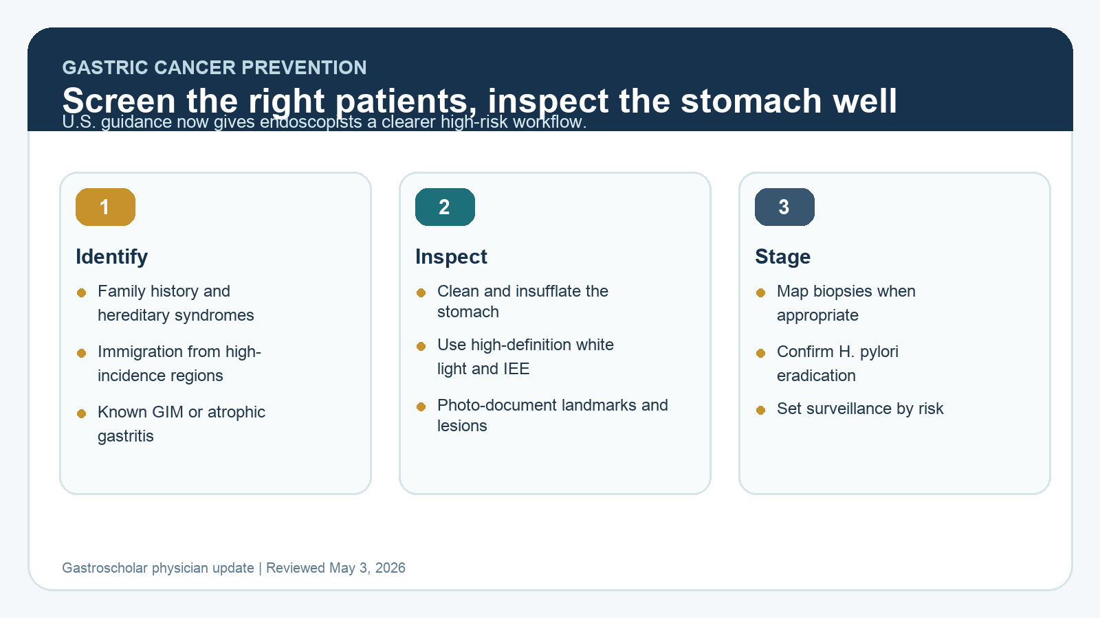
Gastric cancer prevention in U.S. practice is becoming more deliberate. The 2025 AGA update and ACG gastric premalignant conditions guideline do not create population-wide U.S. screening, but they do give gastroenterologists a practical high-risk pathway: identify patients who merit consideration, perform a high-quality upper endoscopy, stage the mucosa when appropriate, eradicate H. pylori, and set surveillance based on endoscopic and histologic risk.
| Question | 2026 Practical Answer | Why It Matters |
|---|---|---|
| Who should be considered? | Patients with family history in a first-degree relative, hereditary syndromes, prior gastric premalignant conditions, or immigration from high-incidence regions may merit screening or surveillance discussion. | Risk is concentrated, so targeted selection is more useful than casual opportunistic biopsying. |
| What test? | High-quality EGD is the key test because it allows inspection, targeted biopsies, and mucosal staging. | Serology or risk scores cannot identify subtle visible neoplasia. |
| What makes the exam high-quality? | Clean mucosa, adequate insufflation, high-definition white light, image enhancement, enough inspection time, photodocumentation, and careful evaluation of the antrum, incisura, body, cardia, and fundus. | Most missed lesions are not missed because no scope was done. They are missed because the stomach was not examined systematically. |
| When are mapping biopsies needed? | Use mapping biopsies when atrophy or gastric intestinal metaplasia needs risk staging, and target any visible lesion separately. | Surveillance decisions depend on extent and severity, not just a yes-or-no pathology result. |
What Changed
The biggest change is that gastric cancer prevention is now framed as a practical U.S. endoscopy quality issue. For years, many U.S. clinicians treated gastric intestinal metaplasia as an incidental pathology phrase with inconsistent follow-up. Current guidance is more structured. Risk selection, mucosal staging, and surveillance decisions should be explicit.
Who Needs a Screening Discussion?
In the United States, gastric cancer risk is not evenly distributed. First-generation immigrants from regions with high gastric cancer incidence, patients with a first-degree family history of gastric cancer, and patients with hereditary cancer or polyposis syndromes deserve a deliberate discussion. Known atrophic gastritis, gastric intestinal metaplasia, dysplasia, autoimmune gastritis, and persistent H. pylori infection also change the threshold for staging and surveillance.
Risk context should be handled carefully. Race, ethnicity, and immigration history are not deterministic biology. They often capture early-life exposure, H. pylori prevalence, dietary and environmental exposures, and access-to-care patterns. The point is not to label patients. The point is to avoid missing preventable gastric cancer in groups where U.S. average-risk assumptions perform poorly.
What the Endoscopist Should Do
A high-quality gastric exam starts before biopsy. Clean mucus and bubbles. Fully insufflate. Slow down in the antrum, incisura, body, cardia, and fundus. Use high-definition white light and image enhancement to inspect for depressed, flat, discolored, or vascular-pattern abnormalities. Photograph landmarks and any suspicious lesion. Target visible abnormalities separately from staging biopsies.
When staging atrophy or gastric intestinal metaplasia, random unlabelled fragments are not enough. A mapped biopsy protocol allows the pathologist and clinician to distinguish limited antral disease from extensive corpus-involving disease. That distinction determines whether surveillance is reasonable and what interval makes sense.
How To Apply This In Practice
- Add a risk prompt to pre-EGD intake: family history, country or region of early life, known GIM or atrophy, autoimmune gastritis, and prior H. pylori.
- Use image enhancement as part of careful inspection, not as a rescue tool after a fast white-light exam.
- Separate targeted biopsies from mapping biopsies in the report and specimen jars.
- Confirm H. pylori eradication when infection is found.
- Document the surveillance rationale rather than writing “repeat EGD per GI.”
Selected Sources
- AGA best practice advice summary for gastric cancer prevention
- ACG Clinical Guideline: Diagnosis and Management of Gastric Premalignant Conditions
Clinical update for gastroenterologists and endoscopists. Reviewed May 2026.