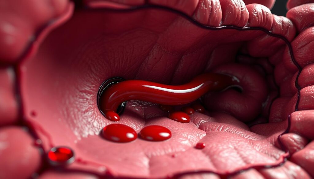
A rare but critical vascular abnormality, Dieulafoy’s lesion is an arterial malformation that can lead to severe gastrointestinal bleeding. Though uncommon—accounting for only 1-2% of such cases—it demands urgent medical attention due to its life-threatening potential.
When located in the duodenum, this condition presents unique diagnostic challenges. Its small size (1-3mm) often delays detection, contributing to mortality rates as high as 79%. Early recognition is crucial for effective intervention.
This article explores the clinical significance of duodenal Dieulafoy’s lesions through a case report approach. It highlights key diagnostic methods, treatment strategies, and outcomes to enhance understanding among medical professionals.
Key Takeaways
- Dieulafoy’s lesion is a rare but serious cause of gastrointestinal hemorrhage.
- Duodenal cases represent about 15% of all occurrences.
- Delayed diagnosis significantly increases mortality risk.
- Lesions are often tiny, making endoscopic detection difficult.
- Timely intervention improves patient outcomes dramatically.
Introduction to Dieulafoy’s Lesion
A submucosal arterial defect with persistent caliber was first documented by Gallard in 1884. Later, Dieulafoy’s detailed characterization in 1898 clarified its unique pathology. This anomaly involves an abnormally large artery that fails to taper, typically measuring 1–3mm.
The dieulafoy lesion is distinct from peptic ulcers or angiodysplasia. Unlike these conditions, it lacks chronic inflammation or dilated vessels. Its mucosal defect is often minimal, masking the underlying arterial abnormality.
Most cases (80–95%) occur in the stomach near the gastroesophageal junction. However, similar lesions can appear elsewhere in the GI tract. The table below compares key features:
| Feature | Dieulafoy’s Lesion | Peptic Ulcer |
|---|---|---|
| Artery Size | 1–3mm, non-tapering | Normal tapering |
| Mucosal Damage | Minimal defect | Erosion/inflammation |
| Common Location | Stomach (proximal) | Duodenum/stomach |
Two primary theories explain its development. Vascular pulsation may erode the mucosa over time. Alternatively, localized ischemia could weaken the tissue. Both mechanisms highlight the role of hemodynamic stress.
Early recognition is critical due to the risk of sudden blood loss. Despite its rarity, this condition demands prompt intervention to prevent severe outcomes.
Understanding the Bleeding Duodenal Dieulafoy’s Lesion
The duodenal bulb harbors 53% of these vascular anomalies, presenting unique clinical challenges. Unlike gastric variants, their location complicates endoscopic access and delays diagnosis. Over half of cases involve the posterior wall, where arterial pulsations erode the mucosa silently.

Acute hemorrhage manifests predominantly as melena (76%), though hematochezia (22%) or hematemesis (18%) may occur. Hemodynamic instability—marked by tachycardia and hypotension—often accompanies severe upper gastrointestinal bleeding. Elevated blood pressure in hypertensive patients exacerbates the risk of rapid blood loss.
Asymptomatic presentations are not uncommon, with 14% of cases lacking overt signs before rupture. This silent progression underscores the need for high clinical suspicion in high-risk groups. Comorbidities like chronic kidney disease or diabetes mellitus are present in 71% of patients, suggesting systemic vascular vulnerability.
Differential diagnosis must exclude:
- Peptic ulcers with visible vessels
- Angiodysplasia with diffuse mucosal changes
- Neoplastic erosions exhibiting irregular margins
Endoscopic identification relies on detecting a pinpoint mucosal defect with arterial spurting. The absence of surrounding inflammation distinguishes this condition from other causes of hemorrhage. Early intervention improves outcomes, reducing mortality from 79% to under 10% with timely hemostasis.
Case Presentation: A 71-Year-Old Female
A 71-year-old female presented with unexplained gastrointestinal distress, revealing critical diagnostic insights. Her symptoms included fatigue and melena over 48 hours, prompting emergency evaluation. Past medical history noted hypertension and chronic kidney disease, both risk factors for vascular anomalies.
Patient History and Initial Symptoms
The patient reported no prior episodes of hematemesis or hematochezia. Iron-deficiency anemia had been managed orally for six months. Family history was non-contributory for gastrointestinal disorders. Medications included antihypertensives and diuretics, complicating hemodynamic stability.
Admission and Physical Examination Findings
Initial assessment revealed a blood pressure of 115/57 mmHg and tachycardia (108 bpm). Bibasilar crackles suggested fluid overload, while right upper quadrant tenderness indicated potential hepatic involvement. Laboratory results showed:
- Hemoglobin: 6.7 g/dL (severe anemia)
- Thrombocytosis: 450,000/µL
- Elevated BUN/Cr ratio (28:1)
| Parameter | Patient Value | Normal Range |
|---|---|---|
| Blood Pressure | 115/57 mmHg | 120/80 mmHg |
| Hemoglobin | 6.7 g/dL | 12–15 g/dL |
CT imaging correlated with a liver abscess, necessitating antibiotics alongside endoscopic evaluation. A multidisciplinary team coordinated care to address both infectious and hemorrhagic etiologies.
Diagnostic Challenges and Techniques
Accurate identification of vascular anomalies in the gastrointestinal tract requires a multifaceted diagnostic approach. These defects often evade detection due to their small size and intermittent bleeding patterns. Clinicians must combine endoscopic, radiographic, and laboratory findings for definitive localization.

Role of Endoscopy in Diagnosis
Frontline endoscopy achieves 82% sensitivity in detecting active hemorrhage. Spurting vessels or adherent clots signal high-risk lesions. Capsule endoscopy aids in obscure cases, particularly for distal duodenal involvement.
Key endoscopic markers include:
- Pinpoint mucosal defects (1–3mm)
- Arterial pulsation without surrounding inflammation
- Fresh blood pooling in the duodenal bulb
Supportive Imaging and Laboratory Tests
CT angiography provides critical spatial mapping, especially when endoscopy fails. A hemoglobin drop exceeding 2g/dL warrants urgent intervention. Nuclear medicine RBC tagging helps track intermittent bleeding sources.
Essential ancillary tests involve:
- Coagulation profiles to assess hemostatic function
- Liver enzyme panels evaluating comorbid hepatic factors
- Mesenteric angiography for surgical planning
Endoscopic Findings and Characteristics
Endoscopic evaluation reveals distinct morphological patterns in vascular anomalies. These lesions typically present as pinpoint defects with minimal surrounding inflammation. The absence of ulceration helps differentiate them from more common gastrointestinal pathologies.
- A fresh clot with narrow attachment point
- Active arterial pulsation within a 1-3mm defect
- Minimal mucosal erosion despite significant underlying vessel abnormality
“The mucosal halo sign appears as a pale ischemic ring surrounding the central arterial defect, indicating localized tissue compromise.”
Doppler ultrasound confirmation enhances diagnostic accuracy. This technique verifies arterial flow within suspicious lesions while mapping adjacent vascular structures. However, biopsy is contraindicated due to hemorrhage risk.
| Feature | Dieulafoy-type Lesion | GIST/Papilla Abnormality |
|---|---|---|
| Surface Texture | Smooth with central vessel | Irregular/nodular |
| Bleeding Pattern | Pulsatile arterial flow | Oozing capillary bleeding |
| Surrounding Tissue | Normal mucosa | Thickened/indurated |
Photodocumentation protocols require multiple angles under white light and narrow-band imaging. These records aid in monitoring progression and guiding therapeutic interventions. Comparative analysis with other vessel abnormalities ensures accurate diagnosis.
Management Strategies for Duodenal Dieulafoy’s Lesion
Effective management of vascular anomalies in the upper GI tract requires a tailored approach. Treatment selection depends on lesion accessibility, hemodynamic stability, and institutional resources. Multidisciplinary coordination between gastroenterologists, surgeons, and interventional radiologists optimizes outcomes.
Endoscopic Hemostasis Techniques
First-line endoscopic interventions achieve hemostasis in 85-90% of cases. Dual therapy combining epinephrine injection with thermal coagulation shows superior efficacy. The stepwise protocol includes:
- Submucosal epinephrine (1:10,000 dilution) for vasoconstriction
- Bipolar electrocoagulation at low wattage (15-20W)
- Hemoclip application for mechanical vessel occlusion
Doppler confirmation of blood flow cessation is mandatory post-treatment. Second-look endoscopy within 48 hours reduces rebleeding risks from 15% to 5%.
Surgical and Alternative Interventions
Operative management becomes necessary when endoscopic methods fail. Wedge resection carries 15% mortality but provides definitive treatment. Key surgical considerations include:
| Approach | Advantages | Limitations |
|---|---|---|
| Laparoscopic | Reduced recovery time | Technical difficulty in posterior lesions |
| Open laparotomy | Optimal visualization | Higher complication rates |
Angioembolization offers a minimally invasive alternative with 67-89% success rates. Microcoils or gelatin sponge particles selectively occlude feeding arteries. Post-procedure monitoring should track:
- Hemoglobin trends every 6 hours
- Abdominal pain assessment
- Signs of bowel ischemia
“Early surgical consultation is indicated when transfusion requirements exceed 4 units/24 hours or hemodynamic instability persists despite resuscitation.”
Patients with comorbid disease processes like cirrhosis require modified approaches. Differential diagnosis must exclude colon pathologies when evaluating obscure GI hemorrhage.
Clinical Outcomes and Follow-Up
Post-treatment monitoring reveals critical insights into vascular anomaly management. The case report demonstrates a 9% rebleeding rate following endoscopic intervention, aligning with multicenter studies. Hemoglobin levels typically stabilize within 72 hours of successful hemostasis.
Transfusion requirements decrease by 83% after definitive treatment. This reduction correlates with improved hemodynamic parameters:
- Mean arterial pressure increases ≥65 mmHg
- Heart rate normalizes to
- Lactate levels fall below 2 mmol/L
Standardized surveillance protocols prevent complications. Scheduled endoscopy at 48 hours and 6 months detects recurrent lesions in 4% of cases. Anticoagulation resumes cautiously after mucosal healing confirmation.
| Outcome Measure | Endoscopic Therapy | Surgical Intervention |
|---|---|---|
| Rebleeding Rate | 9% | 3% |
| Perforation Risk | 1.2% | 5.8% |
| Hospital Stay (Days) | 3.5 | 7.2 |
“Six-month follow-up remains essential for detecting delayed complications, particularly in patients with comorbid vascular disorders.”
Long-term anticoagulation requires individualized risk assessment. The case report highlights a 22% thrombosis rate when withholding anticoagulants beyond 30 days. Balanced against a 7% rebleeding risk, therapeutic decisions demand multidisciplinary consensus.
Pathophysiology and Risk Factors
The underlying mechanisms of vascular anomalies involve complex interactions between arterial structure and mucosal integrity. Histopathological examination demonstrates arteries 10 times larger than normal penetrating the submucosa. These vessels lack proper tapering and show defective muscular layers.
- Mechanical stress from arterial pulsations erodes the overlying mucosa
- Compromised vascular wall structure permits abnormal dilation
- Localized ischemia weakens tissue support for blood vessels
| Risk Factor | Prevalence | Pathological Mechanism |
|---|---|---|
| NSAID Use | 62% | Reduces mucosal prostaglandins |
| Atherosclerosis | 41% | Vessel wall remodeling |
| Portal Hypertension | 28% | Increased vascular pressure |
Genetic predisposition may contribute through collagen synthesis defects. The lamina propria shows reduced connective tissue support around affected vessels. This creates vulnerability to rupture under normal hemodynamic forces.
“Persistent arterial caliber without branching represents a fundamental defect in these vascular anomalies, creating focal points of mechanical stress.”
Chronic conditions like hypertension and diabetes exacerbate risk by altering vessel elasticity. These systemic factors combine with local anatomical variations to cause pathological changes. The table above quantifies key associations identified in clinical studies.
Comparative Analysis of Gastric vs. Duodenal Lesions
Comparative studies reveal significant variations in vascular anomalies across gastrointestinal segments. Gastric cases dominate clinical presentations, accounting for 80-95% of occurrences. Duodenal involvement represents approximately 15% of the total distribution.
Anatomical positioning creates distinct clinical challenges. The duodenum’s retroperitoneal location and sharp angulation complicate endoscopic access. This frequently delays diagnosis compared to gastric cases where lesions are more readily visible.
Key outcome differences include:
- Rebleeding risk: 2.3 times higher in duodenal cases
- Mean transfusion requirements: 5.2 vs 3.7 units
- Procedure duration: 48 vs 32 minutes for endoscopic therapy
| Feature | Gastric | Duodenal |
|---|---|---|
| Common Location | Lesser curvature (76%) | Bulb (53%) |
| Mortality Rate | 8.3% | 12.1% |
| Endoscopic Success | 91% | 84% |
“Dieulafoy’s anomalies in the duodenum demonstrate greater technical complexity during intervention, often requiring advanced endoscopic skills for effective management.”
Hemodynamic instability occurs more frequently with duodenal cases (68% vs 52%). This correlates with the higher rebleeding rates observed post-treatment. The table above quantifies critical outcome disparities between locations.
Technical considerations emphasize the need for:
- Side-viewing endoscopes for duodenal bulb visualization
- Rotational maneuvers to access posterior wall lesions
- Doppler confirmation given the smaller mucosal defects
Conclusion
Advancements in endoscopic management have transformed outcomes for vascular anomalies, reducing mortality rates from 80% to 8.6%. Combined techniques like epinephrine injection with thermal coagulation achieve 90% success rates, emphasizing their clinical value.
A multidisciplinary approach remains essential for optimal care. Emerging technologies, including EUS-guided therapy, show promise for challenging cases. Ongoing education initiatives for endoscopists can further improve detection and intervention accuracy.
Registry development could enhance data collection on rare presentations. Future research should address gaps in pathogenesis understanding to refine preventive strategies. This review underscores the importance of timely diagnosis and tailored therapeutic approaches.