Key Takeaways
- Clinical Bottom Line
- What Changed in the Last 12 Months
- Key Concepts for Endoscopy Leaders
- Patient Selection
Clinical Bottom Line
| Decision Point | Practical Standard | Clinic Implication |
|---|---|---|
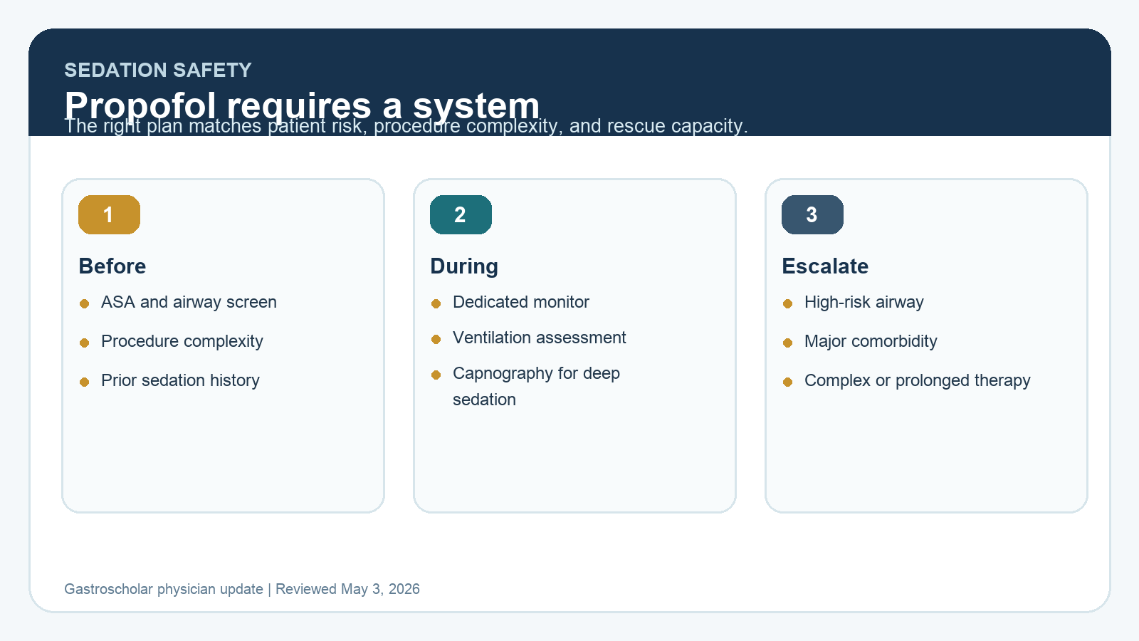
| Endoscopist-directed propofol | Only appropriate inside a credentialed protocol with trained staff, patient selection, and rescue capability. | Treat it as a sedation system, not as a billing workaround. |
| Ventilation monitoring | Capnography should be considered for deep sedation and is required or expected in many anesthesia standards. | Pulse oximetry does not monitor ventilation. |
| Anesthesia involvement | Use anesthesia support for high-risk patients, airway risk, significant comorbidity, and complex or prolonged procedures. | The safest sedation plan is matched to patient risk and procedure complexity. |
What Changed in the Last 12 Months
The core guidance has not flipped. The current update is operational: endoscopy units are under pressure to improve access, control anesthesia costs, and maintain throughput, but sedation shortcuts create patient risk and compliance risk. The article should be read as a protocol checklist for safe implementation, not as an argument that one sedation model fits every room.
ASGE, ASA, and BSG guidance all point in the same direction: sedation is a continuum, ventilation must be assessed, personnel need rescue skills, and deep sedation requires a higher monitoring and staffing standard.
Key Concepts for Endoscopy Leaders
Sedation is a continuum
A patient intended for moderate sedation can drift into deep sedation. A patient intended for deep sedation can require airway support. The unit protocol has to assume that overshoot can happen and define who recognizes it, who rescues it, and when the case stops.
Propofol is not just another benzodiazepine
Propofol gives rapid onset and rapid recovery, but it can produce deep sedation, apnea, hypotension, and loss of airway tone. Endoscopist-directed propofol can be safe in selected settings, but only when local regulation, credentialing, training, patient selection, and dedicated monitoring are aligned.
Capnography changes response time
Pulse oximetry detects oxygenation. Capnography detects ventilation. During deep sedation, capnography can identify apnea or hypoventilation before oxygen saturation falls, especially when supplemental oxygen is being used.
Patient Selection
| Lower-Risk Routine Case | Consider Anesthesia Support |
|---|---|
| ASA I or stable ASA II | ASA III or IV, unstable cardiopulmonary disease, severe pulmonary hypertension, severe valvular disease |
| Normal or low airway risk | Known difficult airway, severe obstructive sleep apnea, high aspiration risk, morbid obesity, limited neck mobility |
| Routine diagnostic EGD or colonoscopy | Complex ERCP, EUS-guided intervention, ESD, POEM, large EMR, prolonged therapeutic case |
| Predictable cooperation and standard positioning | High sedation tolerance, prior sedation failure, prone or semi-prone therapeutic procedure, urgent bleeding with instability |
Minimum Protocol Elements
- Written policy defining which clinicians may administer or direct each sedation level.
- Pre-procedure assessment with ASA class, airway screen, cardiopulmonary risk, aspiration risk, medications, anticoagulation, allergies, and prior sedation history.
- Dedicated monitor who is not simultaneously responsible for noninterruptible technical tasks.
- Vitals, oxygenation, ventilation assessment, level of consciousness, drug dose, and adverse events recorded at defined intervals.
- Immediate availability of suction, oxygen, bag-mask ventilation, airway adjuncts, reversal agents when applicable, defibrillator, emergency drugs, and escalation pathway.
- Recovery criteria that include mental status, ventilation, oxygenation, hemodynamics, pain, nausea, and discharge escort requirements.
Monitoring: What Each Tool Actually Tells You
| Monitor | What It Tells You | What It Misses |
|---|---|---|
| Pulse oximetry | Oxygen saturation | Early hypoventilation, especially with supplemental oxygen |
| Capnography | Breath-by-breath ventilation trend | Does not replace airway assessment or clinical observation |
| Blood pressure | Hemodynamic effect of sedation, bleeding, dehydration, or sepsis | Short apnea events and early obstruction |
| ECG | Rhythm and rate in higher-risk patients | Ventilation and depth of sedation |
| Clinical observation | Airway tone, chest excursion, responsiveness, procedure tolerance | Can lag behind capnography and can be obscured by drapes or positioning |
How to Apply This in Practice
- Separate the sedation decision from the scheduling decision. A fast schedule does not make a patient low-risk.
- Build a pre-procedure triage rule for anesthesia review, including airway risk and complex therapeutic procedures.
- Use capnography for deep sedation and strongly consider it whenever ventilation risk is higher than routine.
- Audit sedation adverse events, airway maneuvers, case interruption, unplanned reversal, EMS transfer, and recovery delay.
- Train the team on jaw thrust, oral and nasal airway placement, bag-mask ventilation, suction, and procedure stop commands.
- Document why anesthesia support was used or not used in borderline cases.
Practice Pitfalls
- Presenting endoscopist-directed propofol as a universal replacement for anesthesia support.
- Relying on pulse oximetry alone during deep sedation.
- Letting the sedation nurse perform continuous monitoring and noninterruptible procedure assistance at the same time.
- Ignoring local regulation, hospital policy, malpractice requirements, or payer rules around propofol.
- Using “routine colonoscopy” language for patients with severe cardiopulmonary disease, difficult airway risk, or prior sedation failure.
Key Sources
- ASGE guideline page: sedation and anesthesia in GI endoscopy
- ASGE sedation and anesthesia guideline PDF
- ASA Standards for Basic Anesthetic Monitoring
- British Society of Gastroenterology sedation guideline
Clinical guidelines summarized by the Gastroscholar Research Team. Last updated: May 3, 2026. This article is intended for physicians and advanced clinicians.