Key Takeaways
- Clinical Bottom Line
- What Changed
- Diagnosis During EGD
- Treatment Options
Clinical Bottom Line
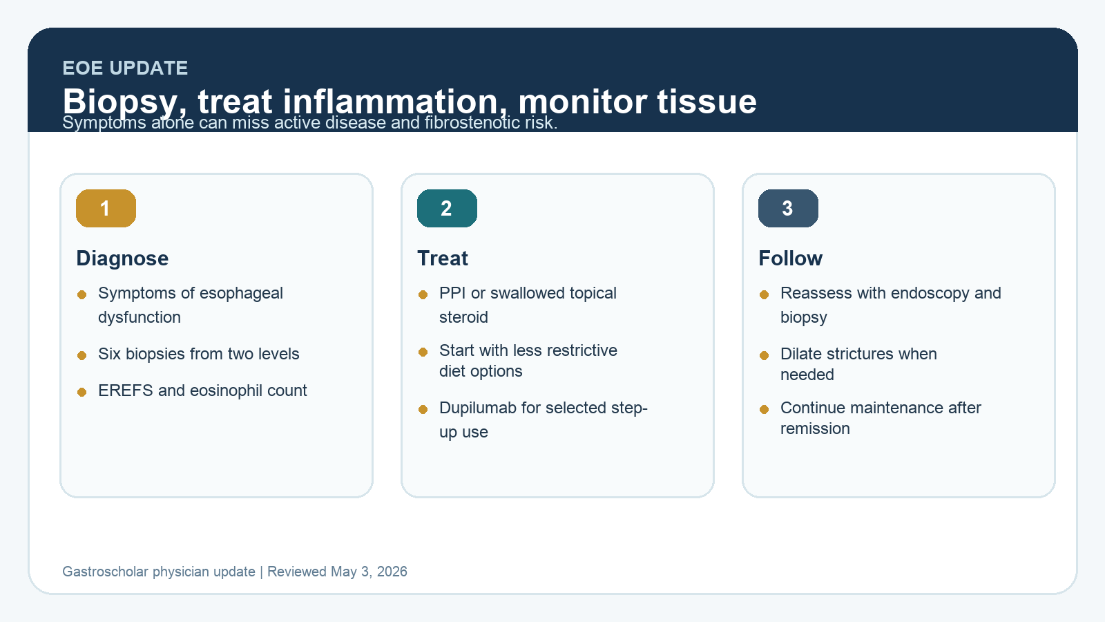
Eosinophilic esophagitis is a chronic inflammatory and fibrostenotic disease. The 2025 ACG update makes the practical workflow clearer: diagnose with symptoms plus esophageal biopsies, document endoscopic phenotype with EREFS, treat inflammation even when dilation is needed, and monitor response with tissue assessment rather than symptoms alone.
| Decision Point | What To Do | Practice Note |
|---|---|---|
| Initial diagnosis | Look for symptoms of esophageal dysfunction and obtain at least 6 targeted biopsies from 2 esophageal levels. | A normal-appearing esophagus does not exclude EoE. |
| Endoscopic description | Use EREFS to document edema, rings, exudates, furrows, and strictures. | Structured scoring makes follow-up exams more useful. |
| Initial therapy | Shared decision-making can start with PPI, swallowed topical steroid, or empiric food elimination. | Less restrictive diets such as 1-food or 2-food elimination are often the most realistic starting point. |
| Fibrostenotic disease | Dilate symptomatic strictures when needed, but pair dilation with anti-inflammatory therapy. | Dilation treats narrowing. It does not treat the underlying inflammation. |
| Monitoring | Reassess symptoms, endoscopic findings, and histology after induction therapy. | Histologic response below 15 eosinophils per high-power field is commonly used as a practical target. |
What Changed
EoE is no longer defined by a failed PPI trial. The modern diagnosis rests on symptoms of esophageal dysfunction, esophageal eosinophilia, and exclusion of other causes. The updated ACG framework also pushes clinicians to stop relying on symptoms alone. Patients can adapt their eating behavior so well that dysphagia appears controlled while active inflammation or stricturing risk persists.
Diagnosis During EGD
The endoscopist should biopsy when EoE is plausible, even if the mucosa looks normal. The practical minimum is 6 targeted biopsies from at least 2 esophageal levels, typically proximal and distal, placed in separate jars when possible. Document rings, edema, exudates, furrows, and strictures with EREFS. This turns a descriptive EGD into a baseline disease assessment that can be compared after treatment.
When clinically feasible, diagnostic endoscopy should be planned so false negatives are less likely. Recent PPI use, swallowed or inhaled steroids, and dietary restriction can reduce visible or histologic activity. That does not mean every medication must be stopped in every patient, but it does mean the endoscopist should know the context before interpreting a borderline biopsy result.
Treatment Options
Initial therapy should be chosen around phenotype, patient preference, adherence, and comorbid atopic disease. PPI therapy remains a reasonable first option for many patients. Swallowed topical steroids, including budesonide or fluticasone formulations, are core anti-inflammatory therapies. Dietary therapy can work, but modern practice generally starts with less restrictive elimination rather than sending every patient directly into a highly restrictive diet.
Dupilumab has become a major step-up option, especially for patients who do not respond to PPI therapy, patients with difficult-to-control disease, or patients with overlapping atopic conditions where a biologic may solve more than one problem. It should not be framed as replacing careful endoscopy. The clinician still needs baseline phenotype, response assessment, and a maintenance plan.
Dilation Still Matters
Fibrostenotic EoE is where the endoscopist’s technical judgment matters most. Dilation can provide meaningful dysphagia relief, especially in narrow-caliber esophagus or focal stricture. The important point is sequencing and framing: dilation addresses the mechanical problem, while anti-inflammatory therapy addresses the disease biology. In many patients, both are needed.
How To Apply This In Practice
- Add an EoE biopsy protocol to dysphagia EGDs, including normal-appearing esophagus when suspicion is present.
- Use EREFS in reports so the next endoscopist can compare disease activity.
- Schedule follow-up endoscopy and biopsies after induction rather than assuming symptom response equals remission.
- For strictures, discuss dilation as symptom therapy and anti-inflammatory treatment as disease control.
- Continue maintenance therapy after remission because EoE is chronic.
Selected Sources
- ACG Clinical Guideline: Diagnosis and Management of Eosinophilic Esophagitis
- ACG Evidence-Based GI summary of the updated EoE guideline
Clinical update for gastroenterologists and endoscopists. Reviewed May 2026.